ADS

Tampilkan postingan dengan label Nursing Care Plan. Tampilkan semua postingan

Kamis, 23 April 2015

NCP for Vomiting - 6 Nursing Diagnosis and Interventions

Nursing Care Plan for Vomiting

Definition

Vomiting is a complex reflex that is mediated by the vomiting center in the medulla oblongata of the brain.

Vomiting is spending gastric contents exclusively through the mouth with the help of contraction of the abdominal muscles. Necessary to distinguish between regurgitation, rumination, or gastroesophageal reflux.
Regurgitation is the food that was issued back-to-mouth due to esophageal peristaltic movement.
Rumination is perpetually conscious of food expenditure to be chewed and then swallowed back.
Gastroesophageal reflux is the return of stomach contents into the esophagus in a passive way that can be caused by hypotonia spingter lower esophagus, abnormal position of the esophagus connection with cardiac or slow emptying of the stomach contents.


Etiology

Discussion of the etiology of vomiting in infants and children by age is as follows:

Age: 0-2 months:

1. Allergic Colitis
Allergy to cow's milk or formula with a soy-based ingredients. Usually followed by diarrhea, rectal bleeding, and cranky.
2. Anatomic abnormalities of the gastrointestinal tract
Congenital anomalies, including stenosis or atresia. Manifestations of food intolerance in the first few days of life.
3. Esophageal Reflux
Regurgitation often occur immediately after feeding. Very often occur in neonates; Clinically important that this situation causes failure to thrive, apnea, or bronchospasm.
4. Increased intracranial pressure
Fussy or lethargy accompanied by abdominal distension, birth trauma and shaken baby syndrome.
5. Malrotation with volvulus
80% of these cases is found in the first month of life, mostly with biliary emesis.
6. Meconium ileus
Inspissated meconium in the distal colon; can be considered a diagnosis of cystic fibrosis.
7. Necrotizing enterocolitis
It often happens, especially in premature babies, especially if experiencing hypoxia at birth. Can be accompanied by irritability or fuss, abdominal distension and hematochezia.
8. Overfeeding
Regurgitation of milk that can not be digested, wet-burps often in infants with excess weight to excess breast milk given.
9. Stenosis pylorus
Peak at the age of 3-6 weeks of life. The ratio of men compared to women is 5: 1 and this situation often occurs in boys first. The clinical manifestations will progressively worsen, projectiles, and non biliary emesis.

Age: 2 months-5 years

1. Brain tumor
Think especially if it is found that the progressive headache, vomiting, ataxia, and no abdominal pain.
2. Diabetic ketoacidosis
Moderate to severe dehydration, a history of polydipsia, polyuria and polyphagia.
3. Corpus alienum
Associated with the incidence of recurrent choking, coughing occurs suddenly or saliva dripping.
4. gastroenteritis
Very often; often their history of contact with sick people, usually followed by diarrhea and fever.
5. Head trauma
Vomiting often or progressive signifies concussion or intracranial hemorrhage.
6. Incarcerated hernia
Onset of crying, anorexia and scrotal swelling that occurs suddenly.
7. Intussusception
The peak occurs at 6-18 months of life; patients rarely experience diarrhea or fever than children who are suffering from gastroenteritis.
8. Posttusive
Often, children will vomit after coughing or coughing repeatedly imposed.
9. pyelonephritis
High fever, looked ill, dysuria or polacisuria. Patients may have a history of urinary tract infections earlier.

Age: 6 years and older

1. Adhesion
Especially after abdominal surgery or peritonitis.
2. Appendicitis
Clinical manifestations and location of pain varies. Symptoms often include increasing pain, radiating to the right lower quadrant, vomiting preceded by pain, anorexia, fever subfebril, and constipation.
3. cholecystitis
More common in women, especially with hemolytic disease (eg, sickle cell anemia). Characterized by epigastric pain or right upper quadrant occurs suddenly after a meal.
4. Hepatitis
Mainly caused by a viral infection or drug-induced; the patient may have a history of bowel movements such as putty colored or tea-colored urine concentrated.
5. Inflammatory bowel disease
Associated with diarrhea, hematochezia, and abdominal pain. Stricture can cause obstruction.
6. Intoxication
More common in children who are learning to walk and adolescents. Suspected if a history of depression. Can also be accompanied by disturbances in mental status.
7. Migraine
Severe headache; often the presence of an aura before an attack such as scotoma. Patients may have a history of chronic headache or a family history of migraine.
8. Pancreatitis
Risk factors include upper abdominal trauma, history of previous infections or moderate infection, corticosteroid use, alcohol and cholelithiasis.
9. Peptic ulcer
In adolescents, the ratio of female: male = 4: 1. Chronic or recurrent epigastric pain, often worse at night time.


Complication

1. Metabolic Complications
Dehydration, metabolic alkalosis, electrolyte and acid-base disorders, depletion of potassium, sodium. Dehydration occurs as a result of fluid loss through vomiting or inputs that are less because of vomiting. Alkalosis as a result of the loss of stomach acid, it is exacerbated by the influx of hydrogen ions into the cell due to potassium deficiency and reduced extracellular sodium. Potassium can be lost along with the material vomit and out through the kidneys together bicarbonate. Sodium can be lost through vomiting and urine. In the state of severe alkalosis, the pH of urine can be 7 or 8, urine levels of sodium and potassium high despite the depletion of sodium and potassium.
2. Failure growth
Repeated vomiting and severe enough cause nutritional disorders due to intake be greatly reduced and when this happens long enough, there will be a failure of growth and development.
3. Aspiration of gastric contents
Material aspiration of vomit can cause asphyxia. Recurrent episodes of mild aspiration cause recurrent respiratory tract infections. This occurs as a consequence of GERD.
4. Mallory Weiss syndrome
A linear laceration on the border of the esophagus and gastric mucosa. Usually occurs in severe vomiting lasts longer. On endoscopic examination found redness of the lower esophageal mucosa LES area. In a short time will heal. When anemia occurs because of heavy bleeding need blood transfusions.
5. Peptic esophagitis
Due to prolonged reflux in chronic vomiting cause mucosal irritation of the esophagus by stomach acid.


Nursing diagnoses that may arise

Fluid volume deficit related to loss of active liquid.
Imbalanced Nutrition: less than body requirements related to absorption disorders.
Nausea related to gastric irritation.
Ineffective tissue perfusion related to hypovolemia.
Risk for Impaired skin integrity related to disruption of metabolic status.
Anxiety related to changes in health status.


Nursing Care Plan for Vomiting

Nursing Diagnosis 1. Fluid volume deficit related to loss of active liquid.
Goal: fluid and electrolyte deficit is resolved.
Expected outcomes:
signs of dehydration: none,
mucosa of the mouth and lips moist, fluid balance.

Intervention:
  • Observation of vital signs.
  • Observation for signs of dehydration.
  • Measure the input and output of fluid (fluid balance).
  • Provide and encourage the family to drink a lot more than 2000 - 2500 cc per day.
  • Collaboration with physicians in fluid therapy, laboratory tests electrolyte.
  • Collaboration with a team of nutrition in low-sodium fluid administration.


Nursing Diagnosis 2. Imbalanced Nutrition: less than body requirements related to absorption disorders.

Goal: nutrients are met.

Intervention:
1. Assess the extent to which the inadequate nutrition clients.
Rational: analyze the causes implement interventions.
2. Estimate / calculate the calorie intake, keep the comments about the appetite to a minimum.
Rationale: Identifying deficiencies / nutritional needs to focus on the problem and create a negative atmosphere affects the input.
3. Measure the weight as indicated.
Rational: Overseeing the effectiveness in diet.
4. Give eat little but often.
Rational: Do not let boredom and nutrient intake can be increased.
5. Encourage oral hygiene before eating.
Rationale: The mouth of the net increase appetite.
6. Offer a drink.
Rationale: It can reduce nausea and relieve gas.
7. consul of a / dislike of patients who cause distress.
Rational: Involve patients in planning, enables patients to have a sense of control and the drive to eat.
8. Provide a varied diet.
Rationale: The food was varied client can increase appetite.
Read More..

Jumat, 05 Desember 2014

Nursing Care Plan for Nasal Cavity Cancer


Nasal Cavity Cancer : 5 Nursing Diagnosis

Nasal Cavity Cancer is cancer that attacks the nasal cavity.

Malignant tumors of the nasal cavity and paranasal sinuses are inseparable because both affect each other.


Causes

The cause of the nasal cavity cancer is not known for certain, but there are some great possibilities, including:
  • Heavy smokers, fistula oroantral, atrophic rhinitis, alkolhol addicts.
  • Chronic infection of the nose and sinuses paranosal.
  • Contact with wood dust on furniture workers (factor of chronic irritation from dust and wood).
  • Contact with industrial materials, such as nickel, chrome, isopropanolol.
  • Thorium dioxide is used as a contrast fluid on X-ray examination.
  • Chronic maxillary sinusitis.


Pathophysiology

Foreign matter (smoke, nicotine, wood dust, nickel, chrome, etc.) into the nasal cavity occurs continuously and for a long time, causing mass formation, changes in the structure and the nasal mucosa, causing obstruction of the nasal cavity to the nasal septum (cavity deformity, nasal septum, trauma cavity / nasal septum, the septum hematoma and septal perforation) or new growth such as nasal polyps, papilloma, inversion and tumor beligna / malignant). In addition, a variety of other reasons cause nasal airway obstruction (adenoid hypertrophy, foreign bodies, atresia, koana, intra-nasal scar tissue, and collapse). This is the mass of the nasal cavity causing edema of the nasal mucosa due to the flow of lymph and venous disorders as well as shaping the polypoid on the nasal cavity. The tumor invades into the upward until cranial fossa and lateral to the orbit.


Signs and Symptoms of Nasal Cavity Cancer

Signs and symptoms of Nasal Cavity Cancer, depending on where the tumor origin and direction as well as the wide-spread.
1. Maxillary sinus tumor and spread to medial.
Signs and symptoms:
  • Nasal congestion.
  • Persistent unilateral rhinorrhea and smelling.
  • Epistaxis.
2. Ethmoid sinus tumors and lamina cribriformis.
Signs and symptoms:
  • Nasal congestion.
  • Anomsia.
  • Runny.
  • Pain frontal area.
3. The basic antrum tumor and extends downward.
Signs and symptoms:
  • Wobbly tooth.
  • Occlusive disorders.
  • Pain in molars.
  • Swelling and lacerations area palate.
4. Tumor extends to the area of the orbit and the nasolacrimal duct.
Signs and symptoms:
  • Diplopia.
  • Proptosis.
  • Blockage of the tear ducts.
  • Eye looks swollen.
  • Musa and orbital palpable.
  • Eyes stand out.
5. Tumor extends to anterior.
Signs and symptoms:
  • Enlargement cheek one side (asymmetric).
6. Advanced stage of the superior alveolar nerve.
Signs and symptoms:
  • Numbness in the upper jaw teeth and gums.
7. The tumor spread and invade into the nasopharynx.
Signs and symptoms:
  • Conductive deafness due to eustachian tube disorders.
8. Another expansion may affect the nerves.
Signs and symptoms:
  • Nerve deafness.
  • Not being able to open the mouth.
  • Facial paresis.
  • Hemiplegia.
  • Hyperesthesia.
  • Severe headache.
  • Changes in eye position.

Nursing Diagnosis for Nasal Cavity Cancer:
  1. Anxiety related to a crisis situation (malignancy), the threat of change in health status-social-economic, function-role changes, changes in social interaction, the threat of death, separation from family.
  2. Disturbance of self-esteem related to deformity of the body due to malignancy, effects of radiotherapy / chemotherapy.
  3. Pain (Acute / Chronic) related to compression / nerve tissue destruction and inflammation.
  4. Imbalanced Nutrition: less than body requirements related to the increase in metabolic status due to malignancy, effects of radiotherapy / chemotherapy and emotional distress.
  5. Risk for infection related to the inadequate secondary defenses and immunosuppressive effects of radiotherapy / chemotherapy.
Read More..

Senin, 24 November 2014

Nursing Care Plan for Gastric Cancer


Definition of Gastric Cancer

Gastric cancer is a malignant form of gastrointestinal neoplasms. Gastric carcinoma is a form of gastric neoplasms are the most common and causes about 2.6 % of all deaths from cancer (Cancer Facts and Figures, 1991).

Gastric cancer occurs in the small curvature or gastric antrum and adenocarcinoma. Other factors, in addition to high- acid foods that cause the incidence of gastric cancer include inflammation of the stomach, pernicious anemia, aclorhidria (no hydrochloride). Gastric ulcer, bacteria H. plylori, and offspring. (Suzanne C. Smeltzer).

Cancer of the stomach or abdominal malignant tumor is an adenocarcinoma. This cancer spreads to the lungs, lymph nodes and liver. Risk factors include chronic atrophic gastritis with intestinal metaplasia pernicious anemia, high alcohol consumption and smoking. (Nettina sandra, nursing practice guidelines).

Gastric cancer is a malignancy that occurs in the stomach, most are of the type adenocarcinoma. Other types of gastric cancer are leiomyosarcoma (smooth muscle cancer) and lymphoma. Gastric cancer is more common in the elderly. Less than 25 % of certain cancers occur in people under the age of 50 years (Osteen, 2003).


Etiology of Gastric Cancer

The exact cause of stomach cancer is unknown, but there are several factors that can increase the development of gastric cancer, include the following matters :

1. Predisposing Factors

a. Genetic factors
Approximately 10 % of patients with gastric cancer have a genetic link. Although still not fully understood, but the mutation of the E - cadherin gene was detected in 50 % of gastric cancer types. The presence of a family history of pernicious anemia and adenomatous polyps was also associated with a genetic condition in gastric cancer. (Bresciani, 2003).

b. Age factor
In this case found to be more common in the age of 50-70 years, but about 5 % of gastric cancer patients aged less than 35 years and 1 % less than 30 years. (Neugut, 1996)


2. Precipitation Factors

a. Consumption of pickled food, smoked or preserved.
Several studies explain the dietary intake of pickled food becomes a major factor increase in gastric cancer. The content of salt that goes into the stomach slows gastric emptying, thereby facilitating the conversion of nitrates into carcinogenic nitrosamines group in the stomach. Combined condition of delayed emptying of stomach acid and an increase in the composition of nitrosamines in the stomach contributed to the formation of gastric cancer (Yarbro, 2005).

b. H.pylori infection.
H. pylori is a bacterium that causes more than 90 % of duodenal ulcers and 80 % of gastric ulcers (Fuccio, 2007). These bacteria on the surface of the gastric ulcer, through the interaction between the bacterial membrane lectins, and specific oligosaccharides from glycoproteins membranes of gastric epithelial cells (Fuccio, 2009).

c. Socioeconomic.
Low socioeconomic conditions are reported to increase the risk of gastric cancer, but not specific.

d. Consume cigarettes and alcohol.
Patients with cigarette consumption of more than 30 cigarettes a day and combined with chronic alcohol consumption increases the risk of gastric cancer (Gonzalez, 2003).

e. NSAIDs.
Inflammatory gastric polyps can occur in patients taking NSAIDs in the long term and this (gastric polyps) may be a precursor of gastric cancer. Gastric polyps conditions will increase the risk of gastric cancer (Houghton, 2006).

f. Pernicious anemia.
This condition is a chronic disease with failure of absorption of cobalamin (vitamin B12), caused by a lack of intrinsic factor gastric secretion. The combination of pernicious anemia with H.pylori infection provides an important contribution to tumorigenesis in the stomach wall formation. (Santacrose, 2008).


Clinical Manifestations of Gastric Cancer

Early symptoms of gastric cancer is often uncertain because most of these tumors in the small curvature, which is only slightly causing interference function of the stomach. Several studies have shown that early symptoms such as pain that is relieved by antacids may resemble symptoms in patients with benign ulcer. Symptoms may include a progressive disease can not eat, anorexia, dyspepsia, weight loss, abdominal pain, constipation, anemia and nausea and vomiting (Harnawati).

Clinical symptoms were found among others (Davey, 2005) :
  • Anemia , vague gastrointestinal bleeding and resulted in an iron deficiency may be a presenting symptom of gastric carcinoma is the most common.
  • Weight loss, common and further illustrates metastatic disease.
  • Vomiting, an indication of the occurrence of gastric outflow obstruction.
  • Dysphagia.
  • Nausea.
  • Weakness.
  • Hematemesis.
  • Regurgitation.
  • Easily satisfied.
  • Enlarged abdominal ascites.
  • Abdominal cramps.
  • Real or faint blood in the stool.
  • The patient complained of discomfort in the stomach, especially after eating.

Complications of Gastric Cancer

1. Perforation

Perforation can occur acute and chronic perforation :
  • Acute Perforation. Perforation often occurs in : ulceration type of cancer that is located in the minor curvature, diantrum near the pylorus. Usually have symptoms similar to perforation of peptic ulcer. These perforations are often found in men (Hadi, 2002).
  • Chronic perforation. Perforations that occur frequently covered by the adjacent tissue, for example by omentum or is penetration. Usually more rare when compared with the complications of benign ulcer. Penetration may be found between layers or layer of omentum gastrohepatic bottom of the liver. What often happens is perforated and covered by the pancreas. With the penetration it will form a fistula, for example gastrohepatic, gastroenteric and gastrocolic fistula. (Hadi , 2002).
2. Haematemesis.
Massive hematemesis and melena occurred ± 5 % of carcinomas ventrikuli whose symptoms are similar as in massive bleeding so much blood is lost, causing hypochromic anemia. (Hadi, 2002).

3. Obstruction.
Can occur in the lower part of the stomach near the pylorus region are accompanied by complaints of vomiting (Hadi, 2002).

4. Adhesion.
If a tumor of the stomach wall can occur adhesion and infiltration of the surrounding organs and cause abdominal pain (Hadi , 2002)


Gastric Cancer - Assessment and 5 Nursing Diagnosis

Nursing Interventions for Gastric Cancer
Read More..

Kamis, 06 November 2014

Nursing Care Plan for Liver Abscess

Anatomy and Physiology of the Liver

The liver is the largest gland in the body, the average weight of 1,500 grams. 2 % of normal weight adults. The liver has two lobes that left and right. Each lobe of the liver is divided into structures called lobules, which is a microscopy unit and functional organ. The human liver has a maximum of 100,000 lobules. Between plates of liver cells are capillaries called sinusoids. Sinusoid limited by phagocytic cells and Kupffer cells. Kupffer cell function is to engulf bacteria and other foreign substances in the blood. (Sylvia a. Price, 2006).

The liver has two sources of blood supply , of the gastrointestinal tract and spleen through the hepatic portal vein and from the aorta through hepatic artery. About one-third of the incoming blood is arterial blood and two-thirds is the portal venous blood. The total volume of blood passing through the liver every minute was 1,500 ml. (Sylvia a. Price, 2006).

The liver is the largest and most important metabolic organ in the body. These organs perform a variety of functions, include the following :
  1. Processing metabolic major nutrients (carbohydrates, fats, proteins) after absorption is the digestive tract.
  2. Detoxification or degradation of residual substances and hormones as well as drugs and other foreign compounds.
  3. Synthesis of plasma proteins, including proteins that are essential for blood clotting, as well as to transport thyroid hormones, steroids and cholesterol in the blood.
  4. Deviations of glycogen, fat, iron, copper, and many vitamins.
  5. The activation of vitamin D.
  6. Spending bacteria from the red blood cells are worn out due to the resident macrophages.
  7. Excretion of cholesterol and bilirubin (Sherwood, 2001)


Definition of Liver Abscess

Liver abscess is a form of infection in the liver caused by a bacterial infection, parasites , fungi and sterile necrosis originating from the gastrointestinal tract characterized by the process of suppuration with the formation of pus in the liver parenchyma (Aru W Sudoyo, 2006).

An abscess is a collection of pus fluid thick, yellowish caused by bacterial, protozoal or fungal invasion into the tissues of the body. Abscesses may occur in the skin, gums, bones, and organs such as the liver, lungs, and even the brain, an area that occurred abscess red and puffy, there is usually a sensation of pain and local heat (Microsoft Encarta Reference Library, 2004).


Causes

Liver abscess is generally divided into two, namely ; amebic liver abscess and pyogenic liver abscess.

a. Amebic liver abscess
Obtained several species of amoeba that can live as a non-pathogenic parasite in the mouth and intestines, but only Entamoeba histolytica that can cause disease. Only some individuals infected with Entamoeba histolytica, which gives the symptoms of invasive, so it is thought there are two types of E. histolytica, namely ; pathogenic and non- pathogenic strain. Variations in the virulence of these strains differ based on its ability to cause lesions in the liver (Aru W Sudoyo , 2006).

E.histolytica in the stool can be found in two forms: vegetative or trophozoite and cyst forms that can survive outside the human body. Mature cyst size 10-20 microns, resistant to dry and acidic atmosphere. Forms tropozoit will die in dry atmosphere and acid. Large trophozoite very active, capable of consuming erythrocytes , which contains protease ; hyaluronidase and mucopolysaccharidase capable of resulting in tissue destruction.

b Pyogenic liver abscess
The infection is mainly caused by gram- negative bacteria and the most common cause is E. coli . Moreover, the cause is Streptococcus faecalis also, Proteus vulgaris, and Salmonellla Typhi. Can also anaerobic bacteria such as Bacteroides, aero bacteria, actinomicosis, and anaerobic streptococci . Necessary for the finding of blood culture, pus, bile, and swabs in anaerobic or aerobic (Aru W Sudoyo, 2006).


Signs and Symptoms

Initial complaint : fever / chills, abdominal pain, anorexia / malaise, nausea / vomiting, weight loss, night sweat, diarrhea, fever (temperature greater than 38°), hepatomegaly, right upper quadrant tenderness, jaundice, ascites, and sepsis the cause of death. (Cameron 1997)

An abscess is the last stage of a tissue infection that begins with a process called inflammation.
Initially, such as bacteria activate the immune system, several events occur :
  1. Blood flow to the area increases.
  2. The temperature of the area increases due to the increased blood supply.
  3. The area swells due to the accumulation of water, blood, and other fluids.
  4. It turns red.
  5. It hurts, because of irritation from the swelling and the chemical activity.
  6. The four signs ; heat, swelling, redness, and pain - the characteristics of inflammation.


Diagnostic Examination

According to Julius, the science of diseases in Volume I, (1998). Investigations among others ;
1. Laboratory
To determine the hematologic abnormalities include hemoglobin, leukocytes, and liver function examination.
2. chest x-ray
Can be found in the form of the right diaphragm, decreased movement of the diaphragm, pleural effusion, lung collapse and lung abscess.
3. Plain abdominal X-ray
Abnormalities may include hepatomegaly, ileus picture, picture of free air above the liver.
4. Ultrasonography
Detecting abnormalities of biliary tract and diaphragm.
5. Tomography
See abnormalities in the posterior and superior , but can not see the integrity of the diaphragm.
6. Serology
Shows a high sensitivity to germs.


Nursing Diagnosis for Liver Abscess

According Doenges, EM (2000), nursing diagnoses of patients with liver abscess include:
  1. Breathing pattern, ineffective related to Neuromuscular, imbalance perceptual / cognitive.
  2. Disturbed Sensory Perception : the process of thought related to chemical changes : the use of pharmaceutical drugs.
  3. Fluid Volume Deficit, Risk for oral fluid intake restriction (process / medical procedure / nausea).
  4. Pain (acute) related to disorders of the skin, tissue, and muscle integrity.
  5. Impaired Skin Integrity related to the interrupt mechanism of the skin / tissue.
  6. Risk for infection related to an operating wounds and invasive procedures.
  7. Disturbed Sleep Pattern related to the disease process, the effects of hospitalization, changes in the environment.
  8. Knowledge deficit (learning need) regarding condition / situation, prognosis, treatment needs.
Read More..

Senin, 13 Oktober 2014

Nursing Care Plan for Esophageal Atresia

Esophageal atresia occurs in about one in 3000-4500 live births, one third of affected children are usually born prematurely. In over 85% of cases, a fistula between the trachea and distal esophageal, accompanying atresia. Very rarely, esophageal atresia or tracheal esophageal fistula occur alone or with a strange combination. Esophageal atresia is a congenital abnormality include group consisting of disruption of continuity of the esophagus with or without connection to the trachea. Infants with Esophageal Atresia is unable to swallow saliva and marked with a bunch of very large amount of saliva and requires suction repeatedly.

The possibility of atresia increased with the discovery of polyhydramnios. Nasogastric tube can still be passed at the time of birth of all babies are born with maternal polyhydramnios as well as infants with excessive mucus, soon after the birth to prove or disprove the diagnosis. In esophageal atresia hose will not pass more than 10 cm from the mouth (confirmation with Rongent chest and abdomen).

Until now not known what teratogenic substances that can cause abnormalities Esophageal Atresia, just reported recurrence rate of about 2% if one of the affected siblings. But now, the theory about the occurrence of esophageal atresia according to most experts no longer associated with a genetic disorder. The debate on embryo-pathological process continues, and only a little is known.

The triggers that cause congenital birth as esophageal atresia are as follows:
  • In the case of polyhydramnios.
  • Preterm infants.
  • If the catheter is used for resuscitation at birth can not enter into the stomach.
There are some circumstances that the symptoms and signs of esophageal atresia, among others:
  • Mouth foaming (bubbles of air from the nose and mouth) and saliva from the mouth of a baby is always melted.
  • Cyanosis.
  • Cough and shortness of breath.
  • Symptoms of pneumonia caused by regurgitation of saliva from the esophagus were clogged and regurgitation of gastric fluid through the fistula into the airway.
  • Abdominal bloating or bulge, because the air through the fistula into the stomach and intestines.
  • Oliguria, because there is no fluid intake.
  • Usually accompanied by other congenital abnormalities, such as heart defects, atresia of the rectum or anus.
  • The presence of aspiration when the baby is drinking.
  • Projectile vomiting.


Nursing Diagnosis for Esophageal Atresia
  1. Impaired swallowing related to mechanical obstruction.
  2. Risk for injury related to surgical procedures.
  3. Anxiety related to difficulty swallowing, discomfort due to surgery.
  4. Altered family processes related to children with physical defects.
Read More..

Minggu, 12 Oktober 2014

Nursing Care Plan for Premature Babies


Newborn infants with gestational age 37 weeks or less at birth is called premature babies. Although small, premature infants in size according to pregnancy, but the development of intra-uterine rudimentary, can cause complications during the post-natal. Newborns whose weight was 2500 grams, or less with a gestational age of more than 37 weeks is called small for gestational age, is different from the premature, although 75 % of neonates whose weight was below 2500 grams born prematurely.

Clinical problems occur more often in premature infants than in full-term infants. Prematurity caused immaturity system development and function, restricting the infant's ability to cope with the problem of disease.

A common problem among others ; respiratory distress syndrome (RDS), necrotizing enterocolitis, hyperbilirubinemia, hypoglycemia, thermoregulation, patent ductus arteriosus (PDA), pulmonary edema, intraventricular hemorrhage. Another additional stressor in infant and parents include hospitalization for illness in infants. Parental responses and coping mechanisms they can cause interference in the relationship between them. Necessary planning and adequate measures for these problems.


Etiology and Precipitating Factors :

Problems in the mother during pregnancy :
  • Diseases / disorders such as hypertension, toxemia, placenta previa, placental abruption, cervical incompetence, multiple fetuses, malnutrition and diabetes mellitus.
  • Low socioeconomic level and inadequate prenatal care.
  • Preterm labor, or induced abortion.
  • Abuse consumption in the mother, such as ; drugs, alcohol, smoking and caffeine.


Assessment
1. History of pregnancy.
2. Status of the newborn.
3. Physical examination head to toe, including : cardiovascular, gastrointestinal, integument, musculoskeletal, neurologic, pulmonary, renal, reproduction.
4. Supporting data
  • X-ray of the chest and other organs to determine the presence of abnormalities.
  • Ultrasonography to detect abnormalities of organs.
  • Stick glucose to determine glucose levels decrease.
  • Serum calcium levels, decreased levels means there is hypocalcemia.
  • Bilirubin levels, to identify improvement (due to premature are more sensitive to hyperbilirubinemia)
  • Electrolyte levels, blood gas analysis, blood type, blood culture, urinalysis, fecal analysis, and so forth.


Nursing Diagnosis

1. Risk for respiratory distress related to immaturity of the lungs, with decreased production surfactan that cause hypoxemia and acidosis.

2. Risk for hypothermia or hyperthermia related to prematurity or changes in ambient temperature.

3. Imbalanced nutrition less than body requirements related to inadequate glycogen reserves, iron, and calcium and loss of glycogen stores due to the high rate of metabolism, inadequate intake of calories, and lose calories.

4. Fluid and Electrolyte imbalances related to immaturity, radiation environment, the effect fototherapy or loss through the skin or lungs.

5. Risk for infection related to immunologic immaturity of the baby and the possibility of infection from mothers or medic / nurse.

6. Risk for impaired skin integrity related to immaturity and fragile skin.

7. Impaired sensory perception : visual, auditory, kinesthetic, gustatory, tactile and olfactory related to less stimulation or excessive in the intensive care environment.

8. Knowledge Deficit (family) about treatment of the sick infant at home.
Read More..

Sabtu, 11 Oktober 2014

Care Plan Examples for Community Health Nursing

Nursing Care Plan - Community Health Nursing


Community is a social group who live in a place, interacting with each other, know each other and have the same interests. Communities are groups of people who live in the same location with the same under the rule, the area or the same location where they live, the social groups that have the same interest.

Health care is a specialized field of nursing which is a combination of science nursing, public health sciences and social sciences are an integral part of the health care provided to individuals, families, groups and communities either healthy or sick comprehensively through promotive, preventive , curative and rehabilitative and resocialization, involving the active participation of the community. Active participation with community health teams are expected to know the health problems faced and solved the problem.

Public health targets are individuals, families / groups and communities, with a focus on primary health efforts, secondary and tertiary. Therefore, public education about the health and social development will help the community in encouraging the spirit to care for themselves, independent living and self-determination in creating optimal health status.

Public participation is required in the individual case. Community as the subject and object of society is able to recognize expected, took the decision to maintain good health. Most of the primary health care goals of society are expected to be able to independently maintain and improve the health status of the community.


Assessment 

Community profile assessment framework (modified)

This assessment is the result of a modification of some previous theories about the community assessment.

Data collection

The data collection is intended to obtain information about health problems in the community to determine which actions should be taken to resolve the issue concerning the physical, psychological, social, economic, spiritual and environmental factors that influence.

Data collection can be done in the following way:

1. Interview or anamnesis

The interview is a mutual communication activities in the form of questions and answers between the nurse with the patient or the patient's family, the community on matters relating to the patient's health problems. Interviews should be conducted with a friendly, open, use simple language and easily understood by the patient or the patient's family, and subsequent interviews or anamnesis recorded in the format of the nursing process.

2. Observations

Observations conducted in community nursing covers aspects of physical, psychological, behavioral and attitude in order to nursing diagnosis. Observations were made using the five senses and the results are recorded in the format of the nursing process.

3. Physical examination

In one community nursing where nursing care is provided nursing care family, the physical examination were performed in an effort to help nursing diagnosis by means of inspection, percussion, auscultation and palpation.


Data processing

Once the data is obtained, the next activity is the processing of data in the following way:
  • Classification of the data or categories of data.
  • Calculation of percentage of coverage.
  • Tabulation of data.
  • Interpretation of the data.

Data analysis

Data analysis is the ability to associate the data with the cognitive abilities possessed that can be known about the gaps or problems faced by the community if it's a problem of health, or nursing problems.

1. Determination of the problem or the formulation of health problems

Based on the analysis of the data can be known health and nursing problems faced by the community, and can be formulated hereinafter intervention. However, the problem has been formulated not to be overcome at once. Therefore we need a priority issue.

2. Priority issues

In determining priority public health issues and nursing need to consider various factors such as the criteria are:
  • Public attention.
  • Prevalence.
  • The severity of the problem.
  • Possible problems to be overcome.
  • Availability of community resources.
  • Political aspects.
Selection or screening community health problems, according to the format Mueke (1988) have screening criteria, among others:
  • In accordance with the role of community nurses.
  • The amount at risk.
  • The magnitude of the risk.
  • The possibilities for health education.
  • The public interest.
  • Possibility to overcome.
  • In accordance with the government program.
  • A resource.
  • Time resources.
  • Funding resources.
  • Equipment resources.
  • Human resources.


Nursing Diagnosis

Nursing diagnosis is the individual's response to health problems whether actual or potential. The actual problem is a problem that is obtained at the time of assessment, while the potential problems are problems that may arise later. So the nursing diagnosis is a statement that is clear, concise and definite about the status and health problems that can be addressed by nursing actions. Thus nursing diagnosis is determined based on problems found. Nursing diagnosis will give an overview of public health issues and the status of both the real (actual), and which may occur.


Nursing Care Plan

Planning nursing is nursing action plans to be implemented to address the problems within their nursing diagnosis has been determined with the aim of fulfilling the needs of the client. So public health nursing care plan is based on pre-defined nursing diagnoses and nursing plan drawn up should include the formulation of objectives, plans nursing actions to be performed and the criteria for assessing the results of the achievement of objectives.

The steps in the planning of public health nursing are as follows:
  • Identify alternative nursing actions.
  • Set techniques and procedures to be used.
  • Involve community participation in planning through village community consultation activities, or mini workshops.
  • Consider the community resources and facilities available.
  • Actions to be implemented must be able to meet the requirement, which was felt by the public.
  • Leads to the objectives to be achieved.
  • Action must be realistic.
  • Arranged sequentially.


References :

(Mubarak, 2005).
(Elisabeth, 2007).
(Riyadi, 2007).
Read More..

Nursing Care Plan for Spinal Cord Tumor

NCP for Spinal Cord Tumor - Assessment, Nursing Diagnosis and Interventions

Spinal cord arranged in the spinal canal and are covered by a layer of connective tissue, dura meter. Spinal cord tumor is an uncommon disorder, and only a few are found in the population. However, if the tumor lesion grows and pressing on the spinal cord, this tumor can cause dysfunction of the limbs, paralysis and loss of sensation.

The incidence of all primary tumors of the spinal cord approximately 10% to 19% of all primary central nervous system tumors. (CNS), and like all tumors in the nervous axis, the incidence increases with age. Gender specific prevalence of almost all the same, except for the meningioma which is generally found in women, and ependymoma are more frequent in males. Approximately 70% of intradural tumors, an extramedullary and 30% is intramedular.

In this case the nurse has an important role in organizing efforts such as improved health (promotion) by way of providing information about the disease, disease prevention (preventive), cure (curative) and rehabilitative.

Complications that can result in spinal cord tumors are very noteworthy because of the impact would worsen the patient's condition, such as; damage the fibers of neurons, loss of sensation of pain (severe circumstances), bleeding metastases, rigidity, weakness, impaired coordination, difficulty urinating or causing loss of control of bladder or constipation.


Definition

Spinal cord tumors are the growth of new tissue in the spinal cord, can be benign or malignant.


Etiology

The pathogenesis of spinal cord neoplasms is unknown, but most arise from abnormal cell growth in the area. Genetic history looks very instrumental in the increased incidence in certain families or syndromic group (neurofibromatosis).


Assessment
  • GCS assessment.
  • Assessment of the level of consciousness.
  • Pathological and physiological reflexes.


Nursing Diagnosis

Diagnosis of tumors of the spinal cord taken based on the results of history and physical examination and investigations. Extradural tumors had a clinical course of spinal cord function will disappear altogether accompanied by spastic weakness and loss of sensation of vibration.

Joint position below the level of the lesion is rapid. On examination of the spine radiogram, most of the patients the tumor will show symptoms of osteoporosis or significant damage to the pedicles and vertebral bodies. Myelogram can confirm the location of the tumor.

In extramedullary tumors, which dominates the symptoms is compression of the spinal nerve fibers, so that the initial look is pain, first in the back and then along the spinal nerve roots. As in the extradural tumors, pain aggravated by traction by movement, coughing, sneezing or straining, and the most severe occurred at night. Pain is intensified at night caused by traction on the nerve root pain, the spine when lengthening after the loss of shortening effect of gravity. Sensory deficits gradually rises to below the level of the spinal cord segments. In extramedullary tumors, CSF protein levels almost always increased. Spinal radiography may show an enlarged foramen and thinning adjacent pedicles. As in the extradural tumors, myelogram, CT scan, and MRI is essential to determine the exact location.

1 Impaired sense of comfort: pain related to increased ICP.

2 Impaired physical mobility related to compression of the blood supply to the corno anterior.

3 Impaired sense of comfort: pain related to intra-thoracic and intradominal.


Nursing Plan

Nursing plan is an action plan that nurses do before nurses perform actions to the patient, who is listed in the nursing plan is:
1. Independent and collaborative interventions, independent intervention is the action to be done independently of nurses to patients, while collaboration is an act of intervention that nurses do in collaboration with other health care team.
2 Criteria of expected results, and
3 Rational, which is the rational benefits of the actions taken by nurses to patients.
Read More..

Rabu, 03 September 2014

Nursing Care Plan for Impaired Swallowing

Impaired Swallowing. Swallowing is a unique process that requires good performance of the muscles in the throat , face, tongue, and palate. The presence of the disease, disorders or abnormalities in one of these organs will interfere with the process of swallowing.

Difficulty swallowing or dysphagia. It is usually a sign of a problem in the throat or esophagus (esophageal), tube-shaped muscle that moves food and liquid from the back of the mouth to the stomach. Although dysphagia can happen to anyone, but generally only occur in the elderly, infants, and those who have problems with the nervous system or brain.

There are many causes that can lead to throat or esophagus does not function normally. It could be because of some small things, but some other things that might cause it is a serious disorder. If only having one or two times only, not to worry, the possibility of not having a medical problem. But if trouble swallowing continuously, it is likely to suffer a serious problem that requires proper handling.

In normal circumstances, the muscles in the throat and esophagus will squeeze or contract to move food and liquids from the mouth to the stomach without obstacles. There are two types of problems that can make it difficult for food and liquid move into the esophagus, namely :

1. The muscles and nerves that help move food through the throat and esophagus does not work properly. This can happen because :
  • Suffered a stroke, brain or spinal injury.
  • Problems with the nervous system, such as post - polio syndrome, multiple sclerosis, muscular dystrophy, or Parkinson's disease as well. It could also be caused after diphtheria, syphilis, poisoning, bibulous, and hysteria.
  • Immune system problems that cause swelling or inflammation, and weakness, such as polymyositis or dermatomyositis.
  • Esophageal spasms. This means that the muscles of the esophagus suddenly pressing. Sometimes this can prevent the food to reach the stomach.
  • Scleroderma. In this condition, the tissues of the esophagus become hard and narrow. Scleroderma can also make the muscles become weak lower esophagus, which can cause food and stomach acid back up into the throat and mouth.
2. There is something blocking the throat or esophagus. This may occur because :
  • Gastroesophageal reflux disease (GERD). When stomach acid up into your esophagus, it can cause ulcers in the esophagus, which then can cause scars or wounds. These scars can make a narrow esophagus.
  • Esophagitis. It is an inflammation of the esophagus. It can be caused by many things, such as GERD or an infection or because the pill is stuck in the esophagus. In addition, difficulty swallowing can also be caused by allergic reactions to certain foods or things other airborne.
  • Diverticula. It is a small sac that grows on the wall of the esophagus or throat.
  • Esophageal tumors. Growth in the esophagus may be cancerous or noncancerous.
  • Lymph nodes and tumors that suppress the esophagus.

In addition , dry mouth could make matters worse dysphagia . This is because you may not have enough saliva to help the food from the mouth to enter the esophagus . Dry mouth can be caused by the influence of the consumption of drugs or other health problems.


Dysphagia can come and go at any time, mild or severe, or worse than would occur continuously. If experiencing dysphagia, may be :
  • Food or liquid could not swallow at swallowing the first experiment.
  • Vomiting, choking, or coughing when swallowing.
  • Food or liquid back up into the throat, mouth, or nose after swallowing.
  • Feeling like food or fluid trapped in one or several parts of the throat or chest.
  • Pain when swallowing.
  • Pain or distress in the chest or stomach.
  • Weight loss due to not getting enough food or fluid intake.

Nursing Care Plan for Dysphagia : Impaired Swallowing
will depend on what is causing dysphagia. Treatment for dysphagia includes :
  • Exercise for the muscles to swallow. If there is a problem with the brain, nerves, or muscles, may need to do exercises to train the muscles to work together to help swallow. Also may need to learn how to good posture or how to put food in the mouth in order to swallow either.
  • Change in eating food. The doctor may suggest to avoid or change certain types of foods and liquids to make the process easier to swallow.
  • Dilation (widening). In this treatment, the device is placed into the esophagus, then carefully will expand the narrow areas of the esophagus. Perhaps this treatment should be done several times.
  • Endoscopy. In some cases, a long, thin scope can be used to retrieve the object that is stuck in the esophagus.
  • Food that is stuck mashed with similar chemicals such as papain, that blob can continue down the food into the stomach.
  • Surgery. If there is something blocking the esophagus (such as a tumor or diverticula), may need surgery to remove it. Surgery is also sometimes used in people who have a problem that affects the muscles of the esophagus (achalasia).
  • Drugs. If experiencing dysphagia associated with GERD, the heat in the stomach, or esophagitis, prescription drugs can help prevent stomach acids enter the esophagus. Infections of the esophagus are often treated with antibiotic drugs.
Read More..

Nursing Care Plan for Dysphagia : Impaired Swallowing

Nursing Diagnosis for Dysphagia -Impaired Swallowing


Swallowing is a complex process that allows the movement of food and liquids from the mouth to the stomach. This process involves structures in the mouth, pharynx, larynx and esophagus.

Complaints of difficulty in swallowing (dysphagia) is one of the symptoms of the disorder or disease in the oropharynx and esophagus. These complaints will arise when there is interference with the movement of the muscles of swallowing and impaired transport of food from the mouth to the stomach. Types of foods that cause dysphagia can provide information regarding disorders that occur.


Purpose

1. General Purpose
  • Knowing nursing care in patients with Dysphagia.
2. Special Purpose
To know the nursing care for patients who experience such as :
  • Definition of Dysphagia.
  • Etiology of Disphagya.
  • Pathophysiology of Disphagya.
  • Diagnosis and treatment Disphagya.
  • Disphagya nursing care to patients.


Nursing Care Plan for Dysphagia


Definition

Disphagya is difficulty in swallowing and getting food from the esophagus into the stomach. Dysphagia can cause all sorts. Important to know the difference dysphagia, because orofaring and esophageal disorders. If not carefully observed, the symptoms are very similar.

On the problems of the esophagus, dysphagia sometimes there is, in the event of esophagitis or esophageal obstruction. Problems of the esophagus is usually also accompanied by regurgitation. Hypersalivation never or rarely occurs and when there is usually a result of a foreign object is actually a pseudo - hypersalivation.


Etiology

Disphagya can be found on some of the causes that can cause the condition include:
  • Stroke.
  • Progressive neurological disease.
  • The tube on trachestomy.
  • Paralise or absence of movement of the vocal cords.
  • Tumors in the mouth.
  • Surgery of the head.


Pathophysiology

Normally people swallow solid food or drinking liquids and swallow saliva or mucus produced by the body hundreds of times every day. The swallowing process has four stages : the first stage of preparation in the mouth, where food or solids mobilized / manipulated and chewed in preparation for swallowing. During the oral stage, the tongue pushing food or solids into the back of the mouth, and began to swallow response. Pharyngeal phase began immediately after food or liquid pass through the pharynx (the tube that connects the mouth to the esophagus) into the esophagus or gastrointestinal tract. The last stage is the stage of esophageal, food or liquid pass through the esophagus into the stomach. Although the first and second stages have some control voluntair, stages three and four occur by itself without realizing it. If the swallowing process stalled due to various reasons, will result in difficulty swallowing.


Nursing Assessment

Nursing assessment needs to be done in patients with swallowing disorders or disphagya include :
  • History of the disease.
  • History of stroke.
  • History of the use of medical devices : tracheostomy, nasogastric tube, mayo tube, ETT, post endoscopy examination.
  • History surgery laryx blood, pharynx, esophagus, thyroid.
  • Postoperative mouth area.

Physical examination :
  • Mouth shape is not symmetrical.
  • Looks an inflammation of the pharynx.
  • The presence of Candida in the oral / mouth.
  • Edema of the pharynx.


Nursing Diagnosis and Nursing Interventions

1. Impaired swallowing related to muscle weakness due to swallowing paralise

Outcomes :
Patients can demonstrate the proper method of swallowing food without causing despair.


Intervention :
a. Review the patient's ability to swallow , note the extent of facial paralysis.
b. Increase efforts to be able to perform effective ingestion such as helping the patient hold his head.
c. Place the patient in a sitting position / upright during and after eating.
d. Stimulation lips to open and close the mouth manually by pressing lightly on the lips / under the chin.
e. Place the food in the mouth is not ill / disturbed.
f. Tap the deepest part of the cheek with a spatula to know the weakness of the tongue.
g. Give eat slowly in a quiet environment.
h. Start by giving a semi-liquid food orally , soft foods when patients can not swallow water.
i. Help the patient to choose foods that are small or do not need to chew and easy to swallow.
j. Instruct the patient to use a straw to drink liquids.
k. Suggest to participate in the exercise program.


2. Imbalanced Nutrition Less than Body Requirements related to lack of adequate food intake.

Outcomes :
Adequate nutritional intake.

Intervention :
a. Instruct the patient to eat slowly and chew food thoroughly.
b. Feeding little and often with foods that are not irritating.
c. Serve food in interesting ways.
d. Avoid eating or drinking foods that contain irritant substances.
e. Measure body weight each day and record the increase.
f. Observation of the patient's intake of nutrients and review the things that hinder / complicate the swallowing.
Read More..

Sabtu, 16 Agustus 2014

5 Diagnosis - NCP for Corneal Ulcer

NCP for Corneal Ulcer
Nursing Care Plan for Corneal Ulcer

Definition

Ulcerative keratitis better known as corneal ulceration, namely the presence of destruction (damage) on the corneal epithelium. (Darling, Vera H, 2000, p 112)


Causes

The reasons include:
  • Abnormalities of the eyelashes (trichiasis) and systems tears (tears insufficiency, lacrimal duct blockage), and so on.
  • External factors, namely: wounds in the cornea (corneal erosio), due to trauma, contact lens use, burns on the face.
  • Corneal abnormalities caused by: chronic corneal edema, keratitis-exposure (on lagophtalmus, general anesthesia, coma); keratitis due to vitamin A deficiency, neuroparalytic keratitis, superficial keratitis virus.
  • Systemic disorders; malnutrition, alcoholism, Stevens-Jhonson, acquired immune deficiency syndrome.
  • Drugs that lower the immune mekaniseme, eg corticosteroids, IUD, local anesthetics and immunosuppressive group.

In etiologic corneal ulcers can be caused by:
  • Bacteria: Germs that can cause corneal ulcers pure is streptokok pneumoniae, whereas other bacterial corneal ulcers caused by trigger factors above.
  • Viruses: herpes simplex, zooster, vaccinia, variola.
  • Fungi: Candida group, Fusarium, Aspergillus, Cephalosporium.
  • Hipersensifitas reaction: The reaction to staphylococcus (marginal ulcers), tuberculosis (keratoconjunctivitis flikten), unknown allergens (ulcers ring). (Sidarta Ilyas, 1998, 57-60)


Pathophysiology
  1. Progressive : In the process of progressive corneal be terihat, infiltration of leukocytes and lymphocytes cells that eat bacteria or necrotic tissue is formed.
  2. Regressive
  3. Establish scarring : In the formation of scar tissue there will be epithelial, new collagen tissue and fibroblasts.

Severity of illness was also determined by the physical state of the patient, a large inoculum and virulence.
Clinical symptoms:
  1. Red eyes.
  2. Mild to severe eye pain.
  3. Photophobia.
  4. Decreased vision.
  5. White opacities in the cornea.

Symptoms that may accompany is the presence of corneal thinning, Descemet folds, corneal tissue reaction (due to interference iris vascularization), a flare, hypopyon, hyphema and posterior synechiae. In corneal ulcers caused by fungi and bacteria are surrounded PMN epithelial defect. When infections caused by viruses, will be seen surrounding hypersensitivity reaction. Usually gram-positive cocci, Staphylococcus aureus and Streptococcus pneumoniae would provide a limited picture of ulcer, round or oval, white gray suppurative ulcers in children. The area that is not exposed cornea will remain clear and no visible color inflammatory cell infiltration. If the peptic ulcer caused by Pseudomonas then be stretched quickly, green yellow purulent material seen attached to the surface of the ulcer.

When ulcers caused by fungi, it will infiltrate surrounded grayed infiltrates surrounding smooth (satellite phenomenon). When the dendrite-shaped ulcer there will be hypesthesia of the cornea. Ulcers can form a fast running descemetocele or corneal perforation which ended by making a form adherent leucoma. When the process of the ulcer is reduced it will show less pain, photophobia, reduced infiltration of ulcers and corneal epithelial defects become increasingly small.


Signs and Symptoms

In ulcers that destroy membranes and stromal bowman, will lead to corneal cicatrix.
Subjective symptoms such as corneal ulcers symptoms of keratitis. Objective symptoms such as ciliary injection, and partial loss of corneal tissue infiltrates. In more severe cases may occur iritis accompanied by hypopyon.
Photophobia.
Pain and lacrimation.

(Darling, Vera H, 2000, p 112)


Diagnostic Examination:
  1. Cards eye / Snellen telebinocular (test visual acuity and central vision)
  2. Tomography measurements: assessing IOP, normal 15-20 mmHg.
  3. Ophthalmoscopy examination.
  4. Blood examination, LED.
  5. EKG.
  6. Glucose tolerance test.


Assessment

  1. Activity / rest: activity changes.
  2. Neurosensory: blurred vision, glare.
  3. Pain: discomfort, pain sudden / severe persistent / pressure in and around eyes.
  4. Security: fear, anxiety.
(Doenges, 2000)


Nursing Diagnosis and Interventions for Corneal Ulcer

1. Fear or anxiety related to sensory impairment and lack of understanding of post-operative care, drug delivery.

Intervention:
  • Assess the degree and duration of visual disturbance.
  • Orient the patient to the new environment.
  • Describe the perioperative routine.
  • Suggest to run the day-to-day living habits when able.
  • Encourage participation of family or people who mean to patient care.

2. Acute pain related to trauma, increased IOP, surgical intervention or administration inflammatory eye drops

Intervention:
  • Give medication to control pain and IOP as prescribed.
  • Give cold compress on demand for blunt trauma.
  • Reduce lighting levels.
  • Encourage the use of sunglasses in strong light.


3. Risk for Self-Care Deficit related to impaired vision.

Intervention:
  • Give instructions to the patient or the person nearest the signs and symptoms, complications should be immediately reported to the doctor.
  • Give oral and written instructions for the patient and the person who means the right techniques in delivering drugs.
  • Evaluation of the need for assistance after discharge.
  • Teach the patient and family guide vision techniques.

4. Disturbed Sensory Perception: Visual related to impaired vision.

Goal: Patient is able to adapt to changes.

Outcomes:
  • Patients receive and resolve in accordance with the limits of vision.
  • Using existing vision or other senses adequately.
Iintervention:
  • Introduce the patient to the environment.
  • Tell patient to optimize other sensing devices that are not impaired.
  • Visit frequently to determine the needs and eliminate anxiety.
  • Involve people in the care and activities nearby.
  • Reduce noise and provide a balanced break.


5. Knowledge Deficit related to lack of information about self-care and disease processes.

Goal: Patients have enough knowledge about the disease.

Outcomes:
  • Patients understand medication instructions.
  • Patients using verbal communication to express the symptoms to be reported.
Intervention:
  • Tell the patient about the disease.
  • Teach self-care during illness.
  • Teach hatching procedure eyedrops and replacement bandage on the patient and family.
  • Discuss the symptoms of the rise in IOP and visual impairment.
Read More..

Rabu, 13 Agustus 2014

Nursing Care Plan for Endophthalmitis

Endophthalmitis - Nursing Assessment and Diagnosis

Definition of endophthalmitis

Endophthalmitis is inflammation of the lining around the inner eye, the fluid in the eyeball (the vitreous humor) and the whites of the eyes (sclera).

Endophthalmitis is a purulent inflammation (suppurative) within the eyeball. Is a purulent inflammation of the entire intra-ocular tissues is accompanied by the formation of abscesses in the body of the glass. Cause of Sepsis, orbital cellulitis, penetrating trauma, ulcer.


Classification

Endophthalmitis can be classified according to:
1 How to enter
  • Endogenous endophthalmitis caused by bacteria spread from elsewhere in the body through the bloodstream. The main fungi. Common predisposing factor is immunocompromised status, septicemia or IV drug abuse.
  • Exogenous endophthalmitis can occur as a result of penetrating trauma or infection in the open surgery eyeball. Endogenous endophthalmitis is very rare, only 2-15% of all endophthalmitis. The main bacteria.

2. Types of agents causing
  • bacteria
  • fungi
  • virus
  • parasites


Etiology

The cause of endophthalmitis among others:
  1. Surgery.
  2. Wounds that penetrate the eye.
  3. Bacteria. The cause of most is Staphylococcus epidermidis, Staphylococcus aureus, and Streptococcus species.
  4. Fungi. The cause of most is Aspergillus, phycomycosis and Actinomyces.


Signs and Symptoms

Inflammation caused by bacteria will provide clinical manifestations of severe pain, red and swollen eyelids, difficult petals opened, chemotic and red conjunctiva, cornea cloudy, cloudy anterior chamber. In addition, there will be a decrease in visual acuity and photophobia (fear of light). Endophthalmitis due to surgery is common after 24 hours and eyesight would worsen with the passage of time. When already deteriorating, will be formed hypopyon, the white fluid-filled sac, in front of the iris.

The symptoms are often severe, which are:
  1. eye pain
  2. redness of the sclera
  3. photophobia (sensitive to light)
  4. visual impairment.

Signs often appear:
  1. eyelids red,
  2. swelling, and difficult to open,
  3. cloudy cornea,
  4. murky chamber of the eye.


Pathophysiology

Endophthalmitis or corpus vitreous abscess is severe inflammation within the eye, usually caused by trauma or surgery, or endogenous due to sepsis. Shaped suppurative inflammation within the eye, and will lead to an abscess in the body of the glass. Exogenous endophthalmitis caused by penetrating trauma or secondary infection following surgery on the open eyeball. Endogenous endophthalmitis caused by the spread of bacteria, fungi or parasites from the focus of infection in the body.
Inflammation by bacteria provide a picture of severe pain, red and swollen eyelids, anterior chamber murky, sometimes accompanied by hypopyon. In the body of the glass can be found masses of white gray and light hippion satellite abscesses form in the body of the glass.


NURSING CONCEPTS

A. Assessment
  • Assessment sharp eyesight.
  • Assessment of pain.
  • Symmetry eyelid.
  • Eye reaction to light / eye movement.
  • Color eyes.
  • The ability to open and close the eyes.
  • Assessment of visual field.
  • Inspect the outside structure of the eye and inspection nodes for the presence of swelling / inflammation.

Data Focus
  • Pain (mild to severe).
  • Photophobia (sensitivity to light) or blepharospasme (eyelid spasms).
  • Sharpness of vision.


Nursing Diagnosis
  1.  Acute Pain: eye related to inflammation and inflammatory processes.
  2.  Disturbed Sensory Perception (specify: visual) related to the inflammatory process.
  3.  Disturbed Body Image related to loss of vision.
  4.  Disturbed Sleep Pattern related to pain.
  5.  Anxiety related to lack of knowledge about the disease.
  6. Knowledge Deficit related to lack of information.
Read More..

Rabu, 16 Juli 2014

Nursing Care Plan for Emphysema - Assessment and Diagnosis

Nursing Care Plan for Emphysema Assessment
Definition of Emphysema

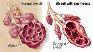
Emphysema is a condition in which the alveoli become stiff expands and continuously filled the air even after expiration. (Kus Irianto.2004.216)

Emphysema is a chronic obstructive disease due to lack of elasticity in the lungs and alveoli surface area. (Corwin.2000.435)


Classification

There are two major types of emphysema, which are classified based on the changes that occur in the lungs:
  1. Panlobular (panacinar), ie damage to the respiratory bronchi, alveolar ducts and alveoli. All air space in the little lobes much enlarged, with little inflammatory disease. The characteristics that have chest hyperinflation, and is characterized by dyspnea on exertion, and weight loss.
  2. Centrilobular (centroacinar), the pathological changes mainly occur in the center of the secondary lobes, and peripheral of acini remain good. Often there is chaos-ventilation perfusion ratio, which lead to hypoxia, hypercapnia (increased CO2 in the arterial blood), polycythemia and heart failure episodes right. The condition leads to cyanosis, peripheral edema, and respiratory failure.


Etiology

Some things that can lead to pulmonary emphysema, namely:
1. Cigarette
Smoking can lead to pathological disorders of the airway ciliary movement, inhibits the function of alveolar macrophages, causing hypertrophy and hyperplasia of bronchial mucous glands.

2. Pollution
Industry and air pollutants can also cause emphysema. The incidence and mortality rates of emphysema can be said to be always higher in areas with high concentrations of industrialization, air pollution as well as tobacco smoke, can cause interference with cilia inhibits the function of alveolar macrophages.

3. Infection
Respiratory tract infections will cause more severe lung damage. Diseases such as respiratory infections, pneumonia, acute bronchiolitis and bronchial asthma, can lead to airway obstruction, which in turn can lead to emphysema.

4. Genetic

5. Exposure to dust


Clinical Manifestations
  • Dyspnea.
  • On inspection: chest shape 'barrel chest'.
  • Chest breathing, abnormal breathing is not effective, and the use of accessory muscles of respiration (sternocleidomastoid).
  • On percussion: hyperresonance and decreased fremitus in all lung fields.
  • On auscultation: audible breath sounds with crackles, and expiratory length.
  • Anorexia, weight loss, and general weakness.
  • Distended neck veins during expiration.


Pathophysiology

Pulmonary emphysema is a lung development, accompanied by tearing of the alveoli that can not be recovered, can be either global or localized, the majority know the whole lung.

Charging excessive air with obstruction, occurs as a result of partial obstruction of the bronchi or bronchioles where the output of the air in the alveoli become more difficult than the input. In such a situation occurs that increases the accumulation of air in the distal alveoli.

In emphysema the narrowing of the airways, it can lead to narrowing of the airway obstruction and tightness, constriction of the airways caused by reduced lung elasticity.


Complication
  • Frequent infections of the respiratory tract.
  • The immune system is less than perfect.
  • The level of lung damage more severe.
  • Chronic inflammatory process in the airways.
  • Pneumonia.
  • Atelaktasis.
  • Pneumothorax.
  • Increase the risk of respiratory failure in patients.


Nursing Assessment  for Emphysema

1. Activity / Rest
Symptoms: Exhaustion, fatigue, malaise, inability to perform daily activities because of difficulty breathing, inability to sleep, need to sleep sitting up high, dyspnea at rest or in response to activity or exercise.
Symptoms: Fatigue, anxiety, insomnia, general weakness / loss of muscle mass.

2. Circulation
Symptoms: Swelling of the lower extremities.
Signs: Increased blood pressure, increased heart rate / severe tachycardia, dysrhythmias, distended neck veins, edema dependent, not associated with heart disease, heart sounds dim (which is associated with increased AP diameter of the chest), color of skin / mucous membranes: normal or gray / cyanosis, pallor may indicate anemia.

3. Foods / Liquids
Symptoms: Nausea / vomiting, poor appetite / anorexia (emphysema), inability to eat due to respiratory distress, permanent weight loss (emphysema), weight gain showed edema (bronchitis).
Signs: poor skin turgor, dependent edema, sweating, drop in body weight, decrease in muscle mass / fat subcutaneously (emphysema), abdominal Palpitations can cause hepatomegaly (bronchitis).

4. Hygiene
Symptoms: Decreased ability / enhancement needs help doing everyday activities.
Signs: Health less, body odor.

5. Respiratory
Symptoms: Shortness of breath (dyspnea hidden emergence as the prominent symptom of emphysema), especially at work, the weather or the recurrence of episodes of difficult airway (asthma), sense of chest pressure, inability to breathe (asthma)
"Air Hunger" chronic.
Shape settled with sputum production every day (especially when awake) for a minimum of 3 consecutive months each year at least 2 years. Sputum production (green, white and yellow) can be a lot of (chronic bronchitis)
Intermittent episodes of cough is usually not productive at an early stage can occur despite earning (emphysema)
A history of recurrent pneumonia: exposure to chemical pollution / respiratory irritants in the long term (eg, cigarette smoke) or dust / smoke (eg, abscess, or coal dust, sawdust)
The use of oxygen at night or continuously.

Signs: Respiratory: usually fast, slow, use of accessory muscles
Chest: hyperinflation with the elevation of the AP diameter, minimal movement of the diaphragm.
Breath sounds: may dim with expiratory wheezing (emphysema); spreads, soft or crackles, wheezing lungs throughout the area.
Percussion: hyperresonant the lung area
Color: pale with cyanotic lips and nail beds.

6. Security
Symptoms History of allergic reaction or are sensitive to substances / environmental factors, presence / recurrence of infection, redness / sweating (asthma).

7. Sexuality
Symptoms: Decreased libido.

8. Social interaction
Symptoms: The relationship of dependence, lack of support systems, improved inability / long illnesses.
Symptoms: Inability to / make maintaining respiratory sounds, physical mobility limitations, abnormalities with the family members.

9. Counseling / Learning
Symptoms: The use / abuse of drugs breathing, difficulty stopping smoking, regular alcohol use, failure to improve.


Nursing Diagnosis  for Emphysema

1. Impaired gas exchange related to ventilation-perfusion abnormalities secondary to hypoventilation.

2. Excess fluid volume related pulmonary edema.
Read More..

Senin, 14 Juli 2014

Guillain-Barre Syndrome Care Plan Nursing


Definition of Guillain-Barre Syndrome

Guillain-Barre Syndrome is an autoimmune disease, in which the immunological process directly on the peripheral nervous system.

Guillain-Barre Syndrome (GBS) is an acute disorder of the nervous system, and diffuse the spinal roots and peripheral nerves and sometimes cranial nerve, which usually occurs after an infection.


Etiology of Guillain-Barre Syndrome

The etiology of Guillain-Barre Syndrome is still not yet known with certainty and is still a matter of debate. Scientists have theorized now is a disorder immunobiology, both in the primary immune response and immune-mediated process. Latent period between infection and symptoms polineuritis gives the notion that there is the possibility of a disorder caused by an allergic reaction in response to peripheral nerve. In many cases, the infection was not previously found, except sometimes the peripheral nerves and spinal ventral and dorsal fibers, there were also disturbances in the spinal cord and medulla oblongata.


Some state / diseases that precede and may have something to do with the occurrence of Guillain-Barre Syndrome, among others:

1. Viral or bacterial infection

Guillain-Barre Syndrome often associated with non-specific acute infection. The incidence of cases of Guillain-Barre syndrome associated with this infection approximately between 56% - 80%, ie 1 to 4 weeks before neurological symptoms arise such as upper respiratory infections or gastrointestinal infection. Acute infection associated with GBS:
a. Viruses: CMV, EBV, HIV, varicella-zoster virus, Vaccinia / smallpox, influenza, measles, mumps, rubella, hepatitis, Coxsackie, Echo.
b. Bacteria: Campylobacter, Jejeni, mycoplasma, Pneumonia, Typhoid, Borrelia B, paratyphoid, brucellosis, Chlamydia, Legionella, Listeria.
2. Vaccination.
3. Surgery, anesthesia.
4. Disease systematic, such as malignancy, systemic lupus erythematosus, thyroiditis, and Addison's disease.
5. Pregnancy or during childbirth.
6. Endocrine disorders.


Clinical Manifestations of Guillain-Barre Syndrome

1. The latent period

The time between infection occurs or circumstances preceding and current prodromal onset of neurological symptoms. The length of the latency period ranging from one to 28 days, an average of 9 days. At this latency period no clinical symptoms arise.

2. Symptoms Clinical

a. Paralysis
The main clinical manifestation is paralysis of the muscles of the lower extremity motor neurone type of limb muscles, body and face sometimes. In most patients, paralysis of both lower extremities begins later spread asenderen to the body, upper limbs and cranial nerves. Sometimes it can also be subject to the four limbs simultaneously, and then spreads to the body and cranial nerves. Paralysis of these muscles symmetrical and followed by hyporeflexia or areflexia. Usually the degree of paralysis of the muscles of the proximal portion of the distal portion is heavier, but it can also be as demanding, or more severe distal part of the proximal portion.

b. Impaired sensibility
Paresthesia is usually more pronounced in the distal extremities, face also may be subject to circumoral distribution. Objective sensory deficit is usually minimal and often with patterns of distribution such as socks and gloves. Exteroceptive sensibility is more commonly known than the proprioceptive sensibility. Muscle pain such as pain often encountered after a physical activity.

c. cranial nerves
Cranial nerves are most commonly known is N.VII. Paralysis of facial muscles often begin on one side but then soon became bilateral, so that the weight could be found between the two sides. All cranial nerves may be subject except N. I and N.VIII. Diplopia could occur from involvement N.IV or N.III. When exposed N.IX and N. X will cause a swallowing difficulty, dysphonia, and in severe cases cause respiratory failure due to paralysis of n. laryngeal.

d. Impaired autonomic function
Impaired autonomic function observed in 25% of patients with GBS. The disorder in the form of sinus tachycardia, sinus bradycardia, or more rarely, so red face (facial flushing), hypertension or hypotension fluctuating, episodic loss of sweating or profuse diaphoresis. Urinary retention or urinary incontinence are rare. This rare autonomic disorder that lasts more than one or two weeks.

e. Respiratory failure
Respiratory failure is a major complication that can be fatal if not treated properly. Respiratory failure is caused by paralysis of the diaphragm and the paralysis of the respiratory muscles, which is found in 10-33 percent of patients.

f. Papilledema
Sometimes encountered papilledema, the cause is not known with certainty. Allegedly due to elevation of the protein content in muscles that cause blockage of fluid arachoidales villi that absorption of cerebrospinal fluid is reduced.


Nursing Diagnosis for Guillain-Barre Syndrome

1. Ineffective breathing pattern
related to:
weakness or paralysis of the respiratory muscles.

2. Disturbed Sensory perception
related to:
changes in reception and transmission.

3. Ineffective Tissue perfusion
related to:
autonomic nervous system dysfunction that causes vascular buildup with decreased venous return.

4. Impaired physical mobility
related to:
neuromuscular damage.

5. Imbalanced Nutrition: less than body requirements
related to:
damage affecting neuromuscular reflex swallowing and GI function.

6. Anxiety
related to:
situational crisis.

7. Pain Acute / Chronic
related to:
neuromuscular damage (paresthesias, disestesia)

8. Knowledge Deficit
related to:
less remembering, cognitive limitations.
Read More..

Jumat, 04 Juli 2014

Care Plan and Nursing Diagnosis for Spina Bifida

Nursing Assessment for Spina Bifida

Subjective and objective data collection on the nervous system disorders, in connection with spina bifida complications depends on other vital organs. Nursing assessment of spina bifida include anamnesis, medical history, physical examination, diagnostic studies, and psychosocial assessment.

1. Anamnesis

The identity of clients includes name, age, gender, education, address, occupation, religion, nationality, date and time of hospital admission, registration number, health insurance, medical diagnostics.

The main complaint is often the reason for a client to ask for help health is the presence of signs and symptoms similar to spinal cord tumors and neurological deficits. Complaints of lumbosacral lipoma on an important sign of spina bifida.


2. History of the disease at this time

Complaints of neurological deficits can manifest as impaired motor (motor paralysis of the lower limbs) and the inferior extremity sensory and / or disorders of the bladder and the sphincter of the stomach. Complaints of unilateral foot deformity and leg muscle weakness is the most common defect. Small feet can occur trophic ulcers and pes cavus. This condition may be accompanied by sensory deficits, especially in the distribution of L3 and S1. Complaints bladder sphincter disorders are found in 25% of infants with neurological involvement, lead to urinary incontinence, urinary dripping, and recurrent urinary tract infections. Usually accompanied by the anal sphincter weakness and sensory disturbance perianal area. Neurological disorders can gradually deteriorate, especially during adolescence mass growth.


3. History of previous illness

Assessment that need to be asked include a history of the growth and development of children, history meningomyelocele ever experienced before, a history of infection subarachnoid space (sometimes chronic or recurrent meningitis), a history of spinal cord tumors, poliomyelitis, spinal developmental disabilities, such as diastematomyelia and foot deformities.


4. Assessment of psychosocial

Assessment of coping mechanisms used and the client's family (parents) to assess the response to illness and changing roles in the family and society as well as responses or influence in their daily lives either in the family or in society. Are there impacts on the client and the parents that raised fears of disability, anxiety, a sense of inability to perform activities optimally.


5. Physical examination

After making the history that led to the complaint the client physical examination is very useful to support the assessment of data from history. Physical examination should be performed by the system (B1-B6) with a focus on examining physical examination B3 (brain) directed and connected with complaints from clients.

a. The general state
In case of spina bifida generally experience loss of consciousness (GCS less than 15), especially if it occurs widely neurological deficits and changes in vital signs.

b. B1 (Breathing)
Changes in the respiratory system associated with inactivity weight. In some circumstances, the results of the physical examination found no abnormalities.

c. B 2 (Blood)
Bradycardia is a sign of changes in brain tissue perfusion. Looked pale skin indicates a decrease in hemoglobin levels in the blood. Hypotension indicates a change in tissue perfusion and early signs of a shock.

d. B3 (Brain)
Spina bifida causes a variety of neurological deficit was primarily due to the effect of increased intracranial pressure. Assessment of B3 (Brain) is a focus and a more complete examination than assessments on other systems.

e. B4 (Bladder)
In the advanced stages of spina bifida, a client may experience urinary incontinence due to confusion and inability to use the urinary system due to damage motor and postural control. Sometimes the external urinary sphincter control is lost or diminished. During this period, intermittent catheterization performed with sterile technique. Urinary incontinence that persists showed extensive neurological damage.

f. B5 (Bowel)
The presence of fecal incontinence that continues to show widespread neurological damage. Bowel examination to assess the presence or absence of bowel sounds and the quality should be assessed prior to abdominal palpation. Bowel sounds are decreased or lost may occur in paralytic ileus and peritonitis.

g. B6 (Bone)
The presence of foot deformity is one important sign of spina bifida. The most common motor dysfunction is the weakness of the lower extremities. To assess the integrity of the skin lesions and sores. Be difficult to move because of weakness, sensory loss or spastic paralysis and fatigue cause problems on the pattern of activity and rest.


6. Diagnostic tests

Spine x-rays to identify any defect in the spine, usually occurs in the posterior arch of the vertebra in the spine midline amount varies. The presence of spinal dyspropism or widening of the spine is a typical sign of radiology at the lumbar (Perkin, 1999).


Nursing Diagnosis for Spina Bifida

1. Urinary incontinence r / t paralysis visceral

2. Risk for injury r / t spastic paralysis

3. Impaired Physical Mobility r / t motor paralysis
Read More..

Acute Pain and Anxiety - NCP for Intestinal Obstruction

Intestinal obstruction (ileus) is a disorder passage of intestinal contents due to blockage resulting in accumulation of fluid and air in the proximal part of the blockage. As a result of the blockage, an increase in intraluminal pressure and intestinal disturbances resorption and increased intestinal secretion. Combined with vomiting as a result of an obstruction or reflux due to regurgitation of stomach full of lead to dehydration, febrile and shock. Obstruction ileus is also an urgency in abdominal surgery is often encountered, is 60-70% of all cases of acute abdomen that is not acute appendicitis. Obstructive ileus also called mechanical ileus.

Based on the mechanism of the obstruction, then the mechanical obstruction can be divided into:
A. Obstruction of the bowel lumen (Intra luminaire), namely:
  • Polypoid tumor.
  • Intussusception.
  • Gallstone ileus.
  • Feces, meconium bezoar (infants).
B. Abnormalities of the intestinal wall (Intramural), mostly congenital in infants:
  • Atresia.
  • Stenosis.
  • Duplication.
In adult patients:
  • Neoplasms.
  • Inflammation.
  • Crohn's disease.
  • Post radiation.
  • Gut connection.
C. Abnormalities outside the colon (Luminaire)
  • Adhesion.
  • External hernia.
  • Neoplasms.
  • Abscess.

Clinical Manifestations : Small Bowel Obstruction

Complaints arising in patients with intestinal obstruction is typical:
  • Abdominal pain, vomiting, obstipation, abdominal distention, no flatus and bowel movement.
  • These painful cramps can be repeated at intervals of 4-5 minutes on intestinal obstruction proximal part. In intestinal obstruction distal part of the frequency increases rarely.
  • After a long obstructed the cramping pain will diminish or disappear because of intestinal distention or movement will be reduced after the strangulation with peritonitis, abdominal pain became severe and continuous.
  • At the proximal intestinal obstruction occurred profuse vomiting with mild distension.
  • At the distal intestinal obstruction, vomiting rarely with vomit the contents of feces, but more severe distension.
  • Increased abdominal circle occurs because of the removal of liquids and gases within the lumen of the intestine due to obstruction in the distal part of the intestine and colon, or paralytic ileus.
  • In the early stages, normal vital signs. Along with the loss of fluid and electrolytes, dehydration will occur with the clinical manifestations of tachycardia and postural hypotension. The body temperature is usually normal but sometimes it can be increased.
  • Physical examination found the presence of fever, tachycardia, hypotension and severe dehydration symptoms.
  • Fever indicates obstruction strangulate. On examination the abdomen appeared distended abdomen obtained and increased peristaltic (sounds borborygmi). In advanced stages where the obstruction continues, peristaltic will weaken and disappear. The presence of feces mixed with blood on rectal examination can toucher suspected malignancy and intussusception.

Nursing Diagnosis : Acute Pain related to an increase in intestinal intraluminal pressure.

characterized by: grimacing expression, complained of feeling pain in the abdominal area.

Goal: expected pain is resolved or controlled.

Outcomes:
  • Revealed a decrease in discomfort.
  • Stating pain at a tolerable level, indicating relaxed.
  • Showed pain control measures.

Intervention:

1) Assess pain with PQRST technique.
Rationale: Monitor and provide an overview of the characteristics of the client and the pain indicators in subsequent interventions.

2) Maintain bed rest in a comfortable position.
Rationale: Bed rest reduces energy use and help control pain and reduce muscle contractions.

3) Teach relaxation or distraction techniques such as listening to music or watching tv.
Rational: to help clients feel more relaxed until the pain can be reduced.

4) Collaboration of analgetic drugs.
Rational: analgesic drugs will block the pain receptors so that pain can not be perceived.


Nursing Diagnosis : Anxiety related to change in health status.

characterized by: increasing the pain of powerlessness, expressed concern.

Goal: expected to decrease anxiety.

Outcomes:

The client will use relaxation techniques to relieve anxiety.

Intervention:
1) Assess the client's level of anxiety.
Rationale: Knowing the coping abilities of individuals.

2) Take time to listen to express anxiety and fear; provide calming.
Rationale: The client will feel better when heard. trusting relationship can be established with the client.

3) Maintain a quiet environment.
Rationale: quiet surroundings make the client more relaxed and can reduce anxiety.

4) Provide diversion through television, radio, games for lowering anxiety.
Rational: to divert the mind from stress and anxiety.

5) Describe the procedures and actions and give an explanation of the strengthening of disease, and prognosis action.
Rationale: patient involvement in care planning can provide a sense of control and helps reduce anxiety.
Read More..

Rabu, 18 Juni 2014

Self-care deficit related to Stroke


Nursing Care Plan for Stroke : Self-care deficit


Definition of Stroke

Stroke is an acute neurological dysfunction caused by impaired blood flow that occur suddenly (within seconds) or raised quickly (within hours) with symptoms and signs corresponding to the focal area disturbed.

Cerebrovascular accident (CVA) also called Stroke is a condition in which the occurrence of neurological deficits caused by decreased blood flow to certain areas of the brain tissue.
Neurological deficits caused by ischemia caused necrotising cells in brain tissue in various areas of the brain.

In the U.S., stroke is the third leading cause of death after heart disease and cancer. This disease can be prevented or minimized by efforts: blood pressure under control, increase awareness of the necessary diet and avoid smoking.


Etiology of Stroke

The occurrence of stroke is caused by the presence of thrombi and emboli that cause the narrowing or occlusion of one of the perfect blood vessels that supply blood to the brain, also if there is bleeding (hemorrhagic). Stroke due to pressure on the walls of blood vessels and arteries spasm, rarely encountered.

1. Thrombosis:

Is the formation of blood clots in blood vessels that can lead to narrowing of the lumen of a blood vessel blockage even happen. Thrombosis is a major cause of cerebral infarction. Two-thirds of strokes are caused by thrombosis due to hypertension and diabetes mellitus both of which can lead to atherosclerosis.

Another factor that can be at risk of thrombosis is an oral contraceptive, coagulation disorders, polycithemia, arteritis, chronic hypoxia, and dehydration. Thrombosis occurs as a result of the formation of atheroma thus narrowing the lumen of blood vessels. Thrombus causing hypoperfusion, infarction and ischemia.

At first occurred paresis (decrease / reduction in force and limb movement), aphasia (language function disorder), paralysis, impaired consciousness, visual disturbances.

2. Embolism:

Blockage / cerebral artery occlusion by an embolus, which resulted in necrosis and edema in the area supplied by the blood vessel blockage.

Embolism is the second leading cause of stroke. Generally derived from the inner lining of the heart (endothelial) where plaque is formed which is then separated and flowed in the blood circulation. If embolism is walking / running on the smaller blood vessels then place it will clog embolism or vascular branching.
Embolism associated with disease / heart problems, namely atrial fibrillation, cardiac infarction, infective endocarditis, rheumatic heart disease, and atrial septal defect. Another cause is not often that air embolism, fat embolism due to fracture femor, amniotic fluid after delivery, and the presence of a tumor.

The attack is sudden. The patient is fully conscious, although patients also feel headache. Prognosis depends location of the blood vessel blockage.

3. Intracerebral hemorrhage:

Bleeding in the brain caused by the rupture of a blood vessel. Intracerebral hemorrhage is usually caused by the presence of hypertension. Another cause is a brain tumor, trauma, thrombolytic treatment, and aneurysm rupture.
Hypertension and atherosclerosis cause degenerative change in the artery walls, causing rupture and hemorrhage. Blood mass will suppress brain tissue. This pressure causes the brain tissue of urgency and decreased blood flow to the brain due to ischemia and infarction.

The area that is often experienced intracerebral hemorrhage putamen and the internal capsule (50%), thalamus, brain hemisper, and pons. Clients will experience a severe headache, nausea and vomiting, loss of ability to walk, dysphagia, eye movement disorders. Bleeding in the post is very dangerous because it is part of the basic life functions. Pons can lead to bleeding in hemiplegia, coma, hyperthermia, and subsequently died.

The prognosis is very bad intracerebral hemorrhage: 70% of patients died due to intracerebral hemorrhage.

4. Subarachnoid hemorrhage:

Caused by the aneurysm, vascular abnormalities, trauma, and hypertension. Aneurysms often occur in patients with atherosclerosis, trauma, hypertension, or vascular abnormalities that are usually congenital bleeding can also be caused by anticoagulant treatment, treatment trhrombolitik, and symphatomimetic.

Bleeding that occurs suppress arachnoid space and cause headache, dizziness, loss of consciousness, nausea, vomiting, fever, pain in the neck and back, paralysis, coma, and later died.


Prevention of Stroke

Primary prevention is to avoid the risk of public health education. Maintain body weight and cholesterol within normal limits, and avoid smoking or using oral contraceptives. Treatment / control diabetes, hypertension and heart disease.

Provide information to clients in connection with the illness with strokes. If already had a stroke, in this situation the goal is to prevent the occurrence of complications with respect to stroke and myocardial wider in the future. In the event of immobility will increase the risk of injury in connection with paralysis and aspiration of the airway. Further Prevention is monitoring the risk factors that can be identified.


Nursing Diagnosis for Stroke : Self-care deficit related to decrease in strength and endurance.

Goal:

Patients can help themselves according to their needs, and be able to express their needs.

Intervention:
  1. Assess the capability and level of deficiency to perform day-to-day needs.
  2. Maintain support, with a strong attitude.
  3. Give positive feedback for any thing done or success.
  4. Avoid doing something for patients to do their own patients, but provide assistance as needed

Rational:
  1. Assist in anticipating / planning meeting individual needs.
  2. Patients will require empathy, care giver to know that will help patients consistently.
  3. Increase feelings of self meaning.
  4. The patient may be very frightened and very dependent.
Read More..