ADS

Tampilkan postingan dengan label Acute Pain. Tampilkan semua postingan

Jumat, 04 Juli 2014

Acute Pain and Anxiety - NCP for Intestinal Obstruction

Intestinal obstruction (ileus) is a disorder passage of intestinal contents due to blockage resulting in accumulation of fluid and air in the proximal part of the blockage. As a result of the blockage, an increase in intraluminal pressure and intestinal disturbances resorption and increased intestinal secretion. Combined with vomiting as a result of an obstruction or reflux due to regurgitation of stomach full of lead to dehydration, febrile and shock. Obstruction ileus is also an urgency in abdominal surgery is often encountered, is 60-70% of all cases of acute abdomen that is not acute appendicitis. Obstructive ileus also called mechanical ileus.

Based on the mechanism of the obstruction, then the mechanical obstruction can be divided into:
A. Obstruction of the bowel lumen (Intra luminaire), namely:
  • Polypoid tumor.
  • Intussusception.
  • Gallstone ileus.
  • Feces, meconium bezoar (infants).
B. Abnormalities of the intestinal wall (Intramural), mostly congenital in infants:
  • Atresia.
  • Stenosis.
  • Duplication.
In adult patients:
  • Neoplasms.
  • Inflammation.
  • Crohn's disease.
  • Post radiation.
  • Gut connection.
C. Abnormalities outside the colon (Luminaire)
  • Adhesion.
  • External hernia.
  • Neoplasms.
  • Abscess.

Clinical Manifestations : Small Bowel Obstruction

Complaints arising in patients with intestinal obstruction is typical:
  • Abdominal pain, vomiting, obstipation, abdominal distention, no flatus and bowel movement.
  • These painful cramps can be repeated at intervals of 4-5 minutes on intestinal obstruction proximal part. In intestinal obstruction distal part of the frequency increases rarely.
  • After a long obstructed the cramping pain will diminish or disappear because of intestinal distention or movement will be reduced after the strangulation with peritonitis, abdominal pain became severe and continuous.
  • At the proximal intestinal obstruction occurred profuse vomiting with mild distension.
  • At the distal intestinal obstruction, vomiting rarely with vomit the contents of feces, but more severe distension.
  • Increased abdominal circle occurs because of the removal of liquids and gases within the lumen of the intestine due to obstruction in the distal part of the intestine and colon, or paralytic ileus.
  • In the early stages, normal vital signs. Along with the loss of fluid and electrolytes, dehydration will occur with the clinical manifestations of tachycardia and postural hypotension. The body temperature is usually normal but sometimes it can be increased.
  • Physical examination found the presence of fever, tachycardia, hypotension and severe dehydration symptoms.
  • Fever indicates obstruction strangulate. On examination the abdomen appeared distended abdomen obtained and increased peristaltic (sounds borborygmi). In advanced stages where the obstruction continues, peristaltic will weaken and disappear. The presence of feces mixed with blood on rectal examination can toucher suspected malignancy and intussusception.

Nursing Diagnosis : Acute Pain related to an increase in intestinal intraluminal pressure.

characterized by: grimacing expression, complained of feeling pain in the abdominal area.

Goal: expected pain is resolved or controlled.

Outcomes:
  • Revealed a decrease in discomfort.
  • Stating pain at a tolerable level, indicating relaxed.
  • Showed pain control measures.

Intervention:

1) Assess pain with PQRST technique.
Rationale: Monitor and provide an overview of the characteristics of the client and the pain indicators in subsequent interventions.

2) Maintain bed rest in a comfortable position.
Rationale: Bed rest reduces energy use and help control pain and reduce muscle contractions.

3) Teach relaxation or distraction techniques such as listening to music or watching tv.
Rational: to help clients feel more relaxed until the pain can be reduced.

4) Collaboration of analgetic drugs.
Rational: analgesic drugs will block the pain receptors so that pain can not be perceived.


Nursing Diagnosis : Anxiety related to change in health status.

characterized by: increasing the pain of powerlessness, expressed concern.

Goal: expected to decrease anxiety.

Outcomes:

The client will use relaxation techniques to relieve anxiety.

Intervention:
1) Assess the client's level of anxiety.
Rationale: Knowing the coping abilities of individuals.

2) Take time to listen to express anxiety and fear; provide calming.
Rationale: The client will feel better when heard. trusting relationship can be established with the client.

3) Maintain a quiet environment.
Rationale: quiet surroundings make the client more relaxed and can reduce anxiety.

4) Provide diversion through television, radio, games for lowering anxiety.
Rational: to divert the mind from stress and anxiety.

5) Describe the procedures and actions and give an explanation of the strengthening of disease, and prognosis action.
Rationale: patient involvement in care planning can provide a sense of control and helps reduce anxiety.
Read More..

Selasa, 29 April 2014

Acute Pain / Chronic Pain - NCP for Rheumatoid Arthritis

Nursing Care Plan for Rheumatoid Arthritis

The changes will occur in the human body in line with the increasing age. Body changes occur early in life until old age in all organs and tissues of the body. Such circumstances also appears on all of the musculoskeletal system and other tissues related to the possibility of some classes of rheumatism. Such circumstances it appears also in all other tissues of the musculoskeletal system and that has to do with the possibility of some classes of Rheumatoid Arthritis. Rheumatoid Arthritis can lead to changes in the muscle, until the function can be decreased when the muscles in the suffering does not trained to activate muscle function. With increasing age old muscle function can be properly trained. But old age is not always experienced or suffered from rheumatism. How the onset of rheumatic this incident, until now has not been fully understood.

According to the consensus of the experts in the field of rheumatology, rheumatism can be expressed as a complaint and / or markings. Of the agreement, otherwise there are three main complaints of the musculoskeletal system are: pain, stiffness and weakness, as well as the three main signs are: swelling of the joints, Muscle weakness, and movement disorders. (Soenarto, 1982).

Rheumatoid arthritis is a long case very often tested. Usually there are many physical signs. Diagnosis of the disease is easily enforced. Management is often a major problem. The incidence of rheumatoid arthritis peaks occur in the fourth decade of age, and the disease is found in women 3 times more often than men. Rheumatoid arthritis is believed to be an immune response to an unknown antigen. Stimulus can be viral or bacterial. There may also predispose to the disease.


Definition

Rheumatoid arthritis is a chronic multisystem disease of unknown cause, is a characteristic feature of rheumatoid arthritis; persistent synovial inflammation, usually of the peripheral joints in a symmetric distribution (Harrison, 2000: 1840).

Rheumatoid arthritis is a chronic disorder that attacks a variety of organ systems, this disease is one of a group of diffuse connective tissue diseases mediated by immune and unknown cause (Sylvia A.Price, 2005: 1385).


Etiology

According to Harrison (2000 : 1841), the cause of rheumatoid arthritis is unknown, it is likely a manifestation of rheumatoid arthritis in response to an infectious agent. One of the possible presence of persistent infection in the joint structure or retention of microbial products in the synovial tissue that trigger chronic inflammatory response. Another potential causative mechanism in rheumatoid arthritis is a disruption of the normal self tolerance which cause reactivity against self antigens in the joints.

But the biggest factor precipitating factors of rheumatoid arthritis is an infection such as bacteria, mycoplasma and viruses (Lemone & Burke, 2001). The main cause of this disorder is unknown. There are several theories put forward as to the cause of rheumatoid arthritis, namely:
  1. Haemolytic streptococcal infection and non-haemolytic streptococci.
  2. Endocrine
  3. Autoimmune
  4. Metabolic
  5. Genetic factors as well as other trigger factors.

At this time, suspected rheumatoid arthritis is caused by autoimmune and infectious factors. This autoimmune reaction against collagen type II; factor for infection may be caused by a virus and mycoplasma organism or group of diphtheroids which produce collagen type II antigens of joint cartilage patients.


Clinical Manifestations

Criteria of the American Rheumatism Association (ARA) 1987 revised are:
  1. Stiff in the morning. Patients feel stiff in the joints and surrounding from waking up at least one hour before maximal improvement.
  2. Arthritis in the three regions. Swelling of soft tissue or bone enlargement joints instead, occurs in at least three joints simultaneously. There are 14 joints that meet the criteria: proximal interphalangeal, metacarpophalang, wrist, elbow, ankle, and metatarsophalangeal left and right.
  3. Artrtis on hand joints. At least there is swelling of the joints of the hands as shown above.
  4. Symmetrical arthritis. That is the involvement of the same joint (not absolutely symmetrical) on both sides simultaneously.
  5. Rheumatoid nodule is a local swelling or tissue lump, usually rather firm to the touch, like an unripe fruit, the which Occurs almost exclusively in association with rheumatoid arthritis.
  6. Serum rheumatoid factor positive. There abnormal titers of serum rheumatoid factor were examined in a way that gives a positive result is less than 5% of the control group.
  7. There is a typical radiological changes on chest X-ray examination postero-anterior hand or wrist, which should indicate the presence of erosions or decalcification of bones located at joints or areas adjacent to joints.
The diagnosis of rheumatoid arthritis is made if at least four of the seven criteria are met above. Criteria 1 till 4 there must be at least 6 weeks.


Nursing Diagnosis for Rheumatoid Arthritis : Pain Acute / Chronic Pain

Acute Pain / Chronic related to the agent of injury, tissue distension by the accumulation of fluid / inflammation, joint destruction.

Outcomes:
Shows the pain is relieved / controlled,
Looks relaxed, sleep / rest and participate in activities according to ability.
Following the prescribed pharmacological programs,
Combining the skills of relaxation and entertainment activities in a pain control program.

Intervention and Rational:
1. Assess pain, note the location and intensity (scale of 0-10). Note the factors that accelerate and signs of non-verbal pain.
R / : Assist in determining the need for pain management and program effectiveness.

2. Provide mat / hard mattress, small pillows. Elevate the patient's bed linens as needed.
R / : Mattress soft / soft, big pillows will prevent the maintenance of proper body alignment, putting stress on diseased joints. Elevation of the bed linens pressure on inflamed joints / pain.

3. Place / monitor the use of pillows, sandbags, rolls trokhanter, splint, brace.
R / :Resting sore joints and maintain a neutral position. The use of the brace can reduce pain and can reduce damage to the joints.

4. Pushes to frequently change positions, help to move in bed, prop joints above and below sick, avoid jerky movements.
R /: Prevent the occurrence of general fatigue and joint stiffness. Stabilize the joint, reducing movement / joint pain

5. Instruct the patient to a warm bath or shower to wake-up time and / or at bedtime. Provide a warm washcloth to compress the affected joints several times a day. Monitor the temperature of the water compresses, bath water, and so on.
R /: Heat improves muscle relaxation, and mobility, decrease pain and stiffness in the morning release. Sensitivity to heat can be removed and dermal wounds can be healed.

6. Provide gentle massage.
R /: Increase relaxation / reduce pain.

7. Encourage use of stress management techniques, such as progressive relaxation, therapeutic touch, biofeedback, visualization, imagination guidance, self hypnosis, and breath control.
R /: Increase relaxation, gives a sense of control and may improve coping skills

8. Involve in entertainment activities appropriate to the individual situation.
R /: Refocus attention, stimulate, and improve self-confidence and feeling healthy.

9. Give medication before activity / exercise planned as directed.
R /: Increase realaksasi, reduce muscle tension / spasm, making it easy to participate in therapy.
Read More..

Senin, 24 Maret 2014

Acute Pain and Bleeding relatde to Ectopic Pregnancy

An ectopic pregnancy is a pregnancy that occurs and is beyond the normal endometrial, endometrial echogenic thickened, as a result of decidual reaction. Uterine cavity is often filled with fluid exudates produced by the decidual cells on examination is seen as anechoic ring structure, called pseudogestational sac.


Signs and symptoms

1. Signs and symptoms of ectopic pregnancy
  • Early pregnancy symptoms (spots or irregular bleeding, nausea, breast enlargement, discoloration of the vagina and cervix, softening the cervix, uterine enlargement)
  • Pain in the abdomen and pelvis.
2. Signs and symptoms of ruptured ectopic pregnancy
  • Collapse and fatigue.
  • The pulse is rapid and weak.
  • Hypotension.
  • Hypovolemia.
  • Acute abdominal pain and pelvic.
  • Abdominal distension.
  • Rebound tenderness.
  • Pale.

Signs and symptoms depend on the length of an ectopic pregnancy, ruptured ectopic pregnancy, abortion, or tubal rupture, gestational age at the time, the degree of bleeding that occurs, and the general state of the patient before pregnancy.

1. Pain.

Pain is the main complaint in ruptured ectopic pregnancy. In tubal rupture, lower abdominal pain occurs suddenly and intensity, accompanied by bleeding that causes people to faint and shock occurs. Usually the tubal abortion, pain is not how great and not continuously. The pain initially located on one side; However, after the entry of blood into the abdominal cavity, pain radiating to the middle or to the entire lower abdomen. Blood in the abdominal cavity can stimulate the diaphragm, causing shoulder pain, and when forming retrouterine hematocele, causing painful defecation.

2. Vaginal bleeding

Vaginal bleeding is an important sign of both the ruptured ectopic pregnancy, suggesting that it is derived from the fetal death and uterine cavity due to the release of the decidua. Bleeding from the uterus and are usually not a lot of dark brown. Bleeding usually occurs around 51-93% on a ruptured ectopic pregnancy. Bleeding means impaired formation of hCG. If there is a solutio placenta decidua can be removed entirely.

3. Amenorrhoea.

Amenorrhoea is the third important sign of ectopic pregnancy, duration of amenorrhea depends on the gestation of the fetus, so it can vary. Most patients do not experience amenorrhea due to fetal death occurred before the next menstruation. Amenorrhea incidence of ectopic pregnancy ranges from 23-97%.

4. On vaginal examination, was found protruding cavity and pain on palpation. Retrouterine hematocele palpable as tumors in douglas cavity, which causes bleeding, and a decrease in blood pressure, increased pulse, and the risk of shock.


Clinical manifestations of ruptured ectopic pregnancy depends on its location. Signs and symptoms vary greatly depending on whether or not the pregnancy ruptured. The symptoms and laboratory test results include:

1. Gastrointestinal complaints.

The most frequent complaints expressed by patients ruptured ectopic pregnancy is pelvic pain. Dorfman stressed the importance of gastrointestinal complaints and vertigo or dizziness. All of these complaints have diversity in terms of incidence due to the speed and extent of bleeding in addition to late diagnosis.

2. Abdominal and pelvic tenderness.

Arising tenderness on palpation of the abdomen and examination, especially by moving the cervix, found in more than three-quarters of cases of ectopic pregnancy or are already experiencing rupture, but it is sometimes not seen before rupture occurrence.

3. Amenorrhea.

History of amenorrhea is not found in a quarter of cases or more. One reason is because patients assume a common vaginal bleeding ectopic pregnancy as a normal menstrual period, thus giving a false date of last menstrual period.

4. Vaginal spotting or bleeding.

During the endocrine functions of the placenta still survive, uterine bleeding usually are not found, but when the endocrine support of the endometrium is no longer sufficient, uterine mucosa will experience bleeding. Bleeding is usually a little, colored dark brown and can be intermittent or persistent.

5. Uterine changes.

Uterus in ectopic pregnancy can be pushed to one side by the ectopic period. In the broad ligament pregnancy or blood filled the broad ligament, the uterus can be shifted great. Uterine cast will be excreted by a minority of patients, perhaps 5% or 10% of patients. Excretion cast fibroids can be accompanied by cramping symptoms similar to networking events abortion spontaneous expulsion of the uterine cavity.

6. Blood pressure and pulse rate.

The initial reaction to the hemorrhage was no change in pulse rate and blood pressure, or sometimes the same reaction as seen in action to be a blood donor phlebotomy is mild increase in blood pressure or vasovagal response accompanied by bradycardia and hypotension.

7. Hypovolemia.
Noticeable drop in blood pressure and pulse rate rise in a sitting position is most often a sign that showed a decrease in blood volume that is quite a lot. All of these changes may have occurred after the onset of serious hypovolemia.

8. Body temperature.
After the acute bleeding, the body temperature may remain normal or even decreased. Higher temperatures rarely found in a state without an infection. Because the heat is a picture that is important to distinguish between who experienced rupture of tubal pregnancy with acute salpingitis, which in this state of body temperatures generally above 38oC.

9. Pelvic mass.
Pelvic mass may be palpable at ± 20 % of patients. The period has the size , consistency and position vary. Usually this mass measuring 5-15 cm , often palpable soft and elastic. However, with the extensive infiltration of the tube wall by the blood of the past can be felt hard. Almost always found in the past pelvic posterior or lateral side of the uterus. Complaints of pain and tenderness often precede palpable in the pelvis future action palpation.
Read More..

Sabtu, 15 Maret 2014

Acute Pain - Nursing Diagnosis and Interventions for Urolithiasis

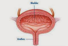
Kidney stones in the urinary tract (urinary calculus) is hard as a rock mass formed in the urinary tract and can cause pain, bleeding, infection or blockage of urine flow.

These stones can form in the kidneys (kidney stones) and in the bladder (bladder stones).
The process of stone formation is called urolithiasis (renal lithiasis, nephrolithiasis). The concentration of stone-forming substances high in blood and urine as well as eating habits or certain medications, can also stimulate the formation of stones. Anything that impedes the flow of urine and cause stasis (no movement) in the urine anywhere in the urinary tract, increasing the likelihood of stone formation.

Stone, especially small ones, may not cause symptoms. Stone in the bladder can cause pain in the lower abdomen. Stones that obstruct the ureter, renal pelvis and renal tubules can cause back pain or renal colic (severe colicky pain). Renal colic is characterized by severe pain intermittent, usually in the area between the ribs and hip bones, which spread to the abdomen,  pubic area and inner thighs. Other symptoms are nausea and vomiting, abdominal distention, fever, chills and blood in the urine. Patients may be frequent urination, especially when the stone passes through the ureter. Stones can cause urinary tract infections. If stones block the flow of urine, the bacteria will be trapped in the urine collected over the blockage, so that there was an infection. If the blockage lasts long, the water will flow back into the urinary tract in the kidney, leading to suppression of which would inflate the kidneys (hydronephrosis) and eventually kidney damage can occur.

Common symptoms of kidney stone disease are:
  • Urinate more often occurs
  • Pain at the waist
  • Sometimes accompanied by fever and seizures
  • Cloudy yellow urine
  • A history of kidney stones who previously suffered by one member of the family

Nursing Diagnosis for Urolithiasis : Acute Pain related to increase in the frequency of ureteral contractions, tissue trauma, edema and cellular ischemia.

Nursing Interventions:

1. Record the location, duration / intensity of pain (scale 1-10) and its spread. Pay attention to non-verbal signs such as: increase in BP and pulse rate, restlessness, grimacing, moaning, floundering.

2. Explain the causes of pain and the importance of reporting to the nursing staff of any changes that occur pain characteristics.

3. Perform actions that promote comfort (such as light massage / warm compress on the back, quiet environment)

4. Help the patient to deep breathing, guided imagery and therapeutic activity.

5. Help / encourage increased activity (ambulation active) as indicated with at least 3-4 liters of fluid intake per day within cardiac tolerance.

6. Note the increase / persistence of abdominal pain.

7. Collaboration of appropriate drug therapy program.

8. Maintain urinary catheter patency when needed.


Rational:

1. Help evaluate the progress of obstruction and stone movement. Pelvic pain often spreads to the back, groin, genitalia with respect to proximity plexus nerves and blood vessels that supply the other areas. Sudden pain and can lead to severe anxiety, fear / anxiety.

2. Reported early pain, analgesic provision provides an opportunity at the right time and help improve the client's coping ability in reducing anxiety.

3. Promote relaxation and reduce muscle tension.

4. Divert attention and help to relax the muscles.

5. Physical activity and adequate hydration increases the passage of the stone, prevent urinary stasis and prevent further stone formation.

6. Complete obstruction of the ureter may lead to perforation and ekstravasasiurine into perrenal area, this is an acute surgical emergency.

7. Prevent stasis / urinary retention, lowering the risk of increased pressure and kidney infections.
Read More..

Rabu, 22 Januari 2014

Acute Pain and Anxiety - NCP for Bladder Cancer

Bladder cancer is a cancer of the bladder organ. Bladder is the organ that serves to accommodate the urine from the kidneys. If the bladder is full of urine then it will be removed.

The exact cause of bladder cancer is not known. But studies have shown that these cancers have multiple risk factors, namely:
  • Age, the risk of bladder increases with age.
  • Smoking is a major risk factor.
  • Work environment, some workers have a higher risk of developing this cancer because of its place works found carcinogenic substances (cancer-causing).
  • Race, white people have a 2 times greater risk, there is the smallest risk among Asians.
  • Men, are at risk 2 - 3kali greater.
  • Family history, people whose family is suffering from bladder cancer have a higher risk of developing this cancer. Researchers are studying the change of certain genes that may increase the risk of this cancer.

Nursing Diagnosis and Interventions for Bladder Cancer

1. Acute Pain
related to:
  • disease process (suppression / destruction of nerve tissue, nerve supply system infiltration, nerve pathway obstruction, inflammation),
  • side effects of cancer therapy
characterized by:
  • clients say pain,
  • clients have difficulty sleeping,
  • not able to focus, expressions of pain, weakness.
Goal:
  • Clients are able to control pain through activity.
  • Reported experiencing pain.
  • Following treatment program.
  • Demonstrate techniques of relaxation and diversion of pain through activity.
Interventions:
  • Determine history of pain, location, duration and intensity.
  • Evaluation of therapy: surgery, radiation, chemotherapy, biotherapy, teach the client and family about how to deal with.
  • Give diversion such as repositioning and fun activities such as listening to music or watching TV.
  • Encourage stress management techniques (relaxation techniques, visualization, guidance), happy, and provide therapeutic touch.
  • Evaluation of pain, provide treatment if necessary.
  • Discuss pain management with doctor and also with clients.
  • Give analgesics as indicated.

Rational:
  • Provide the necessary information for planning care.
  • To determine the appropriate therapy is carried out or not, or even cause complications.
  • To improve the comfort of the clients distract from pain.
  • Improving self-control over side effects by lowering stress and anxiety.
  • To determine the effectiveness of pain management, pain levels and to the extent the client is able to withstand, and to investigate the needs of the client will be anti-pain medication.
  • In order for a given targeted therapy.
  • To cope with the pain.


2. Anxiety
related to:
  • crisis situations (cancer),
  • changes in health,
  • socio-economic,
  • roles and functions,
  • forms of interaction,
  • preparation for death,
  • separation of the family

characterized by:
  • increase in tension,
  • fatigue,
  • express awkwardness role,
  • feeling dependent,
  • inadequate ability to help themselves,
  • sympathetic stimulation.
Goal:
  • Clients can relieve anxiety.
  • Relax and be able to see themselves objectively.
  • Demonstrate effective coping and able to participate in treatment.
Interventions:
  • Determine the client's previous experience of the illness.
  • Provide accurate information about prognosis.
  • Give the client a chance to express anger, fear, confrontation. Give the information with reasonable emotions and expressions appropriate.
  • Explain the treatment, the purpose and side effects. Help clients prepare for the treatment.
  • Record ineffective coping as less social interaction, lack of empowerment, etc..
  • Encourage to develop interaction with the support system.
  • Provide a quiet and comfortable environment.
  • Maintain contact with clients, talk and touch with the fair.
Rational:
  • Data about previous client experience will provide a basis for extension and avoid duplication.
  • Provision of information to assist clients in understanding the disease process.
  • Can reduce client anxiety.
  • Assist the client in understanding the need for treatment and side effects.
  • Knowing and explore coping patterns and handle client / provide solutions in an effort to improve the strength in overcoming anxiety.
  • So that clients receive support from the closest person / family.
  • Give the client a chance to think / contemplate / break.
  • Clients gain confidence and belief that he really helped.
Read More..

Rabu, 15 Januari 2014

Acute Pain and Fatigue - NCP for Systemic Lupus Erythematosus

Nursing Care Plan for Systemic Lupus Erythematosus

SLE (Systemic Lupus Erythematosus) is an autoimmune condition that affects multiple organ systems. Its pathology is related to the release of antibodies that bind to normal nuclear components. Lupus can attack any organ and system in the body. For unknown reasons, in systemic lupus erythematosus, the body forms auto-antibodies against these normal molecules.

The signs and symptoms of lupus may occur rapidly or develop slowly. They may be either mild or severe and may be either temporary or permanent. Most people with lupus will experience episodes or "flares". This is simply where the signs and symptoms get worse or they can improve or even disappear completely for a period of time.


Nursing Diagnosis and Interventions 

1. Acute Pain related to inflammation and tissue damage.

Goal: improvement in comfort level

Intervention:
  1. Carry out a number of actions that provide comfort (heat / cold; massage, position changes, break; foam mattresses, pillows buffer, splints; relaxation techniques, activity that distracts)
  2. Provide anti-inflammatory preparations, analgesics as recommended.
  3. Adjust treatment schedule to meet the needs of patients to pain management.
  4. Encourage the patient to express his feelings about the nature of chronic pain and illness.
  5. Describe the pathophysiology of pain and helping patients to realize that pain is often brought him to the method of unproven therapies.
  6. Assist in identifying a person's life that brings pain to the patient cases using unproven therapies.
  7. Perform an assessment of the subjective changes in pain.

2. Fatigue related to an increase in disease activity, pain, depression.

Goal: include action as part of the activities of daily living necessary for change.

Intervention:
1. Give an explanation of fatigue:
  • The relationship between disease activity and fatigue.
  • Explain the actions to provide comfort while executing.
  • Develop and maintain a sleep routine actions fatherly (warm water bath and relaxation techniques that facilitate sleep).
  • Explaining the importance of rest to reduce systemic stress, articular and emotional.
  • Explains how to use traditional techniques to save energy.
  • Identify the factors that lead to physical and emotional exhaustion.
2. Facilitating the development schedule of the activity / rest right.
3. Encourage patients' adherence to treatment programs.
4. Refer and thrust conditioning program.
5. Encourage adequate nutrition including iron from food sources and supplements.
Read More..

Minggu, 12 Januari 2014

Acute Pain and Impaired Physical Mobility NCP for Tuberculous Meningitis

Tuberculosis meningitis is a TB infection of the brain and the spinal cord. The initial symptoms can be irritability and restlessness. Later the patient may develop other symptoms such as a stiff neck, headaches, vomiting, variations in mental behaviour, seizures, or coma.

Nursing Care Plan for Tuberculous Meningitis

Nursing Diagnosis I :

Acute pain related to the process of infection in the central nervous system

Goal:
1. Long-term goal
Pain is gone.

2. Short-term goals
The pain gradually diminished

Outcomes:
  • Clients reported no pain, or pain can be controlled.
  • Shows posture relaxed and able to sleep / rest appropriately.
Intervention
1. Provide a quiet environment, the room is rather dark as indicated.
rational:
Lowering the reaction to outside stimulation or sensitivity to light and improve the rest / relaxation.

2. Put an ice bag on head, clothes on cold eyes.
rational:
Increases vasoconstriction, blunting sensory perception which will further decrease the pain.

3. Support to find a comfortable position, such as head a little bit higher.
rational:
Lowering of meningeal irritation, discomfort resultant further.

4. Give range of motion exercises active / passive appropriately and do massase muscular shoulder or neck area.
rational:
Can help relax the muscle tension that increases the reduction of pain or discomfort.


Nursing Diagnosis II :

Impaired physical mobility related to neuromuscular damage

Goal:
1. Long-term goal
Physical mobility increased / improved

2. Short-term goals
Impaired physical mobility gradually decreased

Outcomes:
Client is able to mobilize.

Intervention
1. Check back ability and the functional state of the damage.
rational :
Identify possible damage affecting functionally and intervention options that will be done.

2. Assess the degree of immobilization of the client by using the scale dependence.
rational :
The client is able to self (value 0) or need help / tools are minimal (score 1) ; need help being supervised / taught (score 2) ; need help / tools that continuously and special tools (value 3) , or depending on the total the provision of care (Grade 4) ; someone in all categories are equally at risk of accidents , but the category with a value of 2-4 has the greatest risk for the occurrence of such hazards in connection with immobilization.

3. Give or aids to perform range of motion exercises / ROM.
rational :
Mobilization and maintain joint function / normal position and reduce the occurrence of venous limb static.

4. Provide meticulous skin care, massage with moisturizer and change linen / clothes wet and keep the linens are kept clean and free of wrinkles.
rational :
Improves circulation and skin elasticity and reduce the risk of skin excoriation.
Read More..

Rabu, 21 Desember 2011

Nursing Diagnosis of Acute Pain related to Constipation

Nursing Diagnosis of Acute Pain related to Constipation


Acute pain related to the accumulation of hard stool in the abdomen

Goal: show pain has been reduced

Expected Outcomes:
  • Relaxation techniques individually demonstrate effective to achieve comfort
  • Maintaining the level of pain on a small scale
  • Reported physical and psychological health
  • Recognize factors and using measures to prevent pain
  • Using action to reduce the pain with analgesics and non-analgesic appropriately
Nursing Interventions Acute Pain related to Constipation

1. Help the patient to focus more on the activity of the pain by doing penggalihan through television or radio.
Rationale: The client can distract from pain.

2. Note that the elderly have increased sensitivity to the analgesic effects of opiates.
Rational: Be careful in giving anlgesik opiates.

3. Consider the possibility of drug interactions in the elderly.
Rational: Be careful in the provision of drugs in the elderly.

4. Ask the patient to assess pain or lack of comfort on a scale of 0-10
Rationale: Knowing the client's level of perceived pain

5. Use the pain flow sheet
Rationale: Knowing the characteristics of pain

6. Perform a comprehensive pain assessment
Rational: In order for the specific pain mngetahui

7. Instruct patient to meminformasikan on nurses if the pain-reducing achieved less
Rationale: Nurses can perform appropriate action in addressing the client's pain

8. Give pain neighbor information
Rational: In order for the patient does not feel anxious.
Read More..